
Case contribution: Dr Radhiana Hassan
Clinical:
- A 48 years old female
- Presented with dizziness and vertigo, no tinnitus, no hearing disturbance
- No loss of consciousness, no seizure, no headache or blurred vision
- Clinically cranial nerves are intact. No nystagmus.
MRI findings:
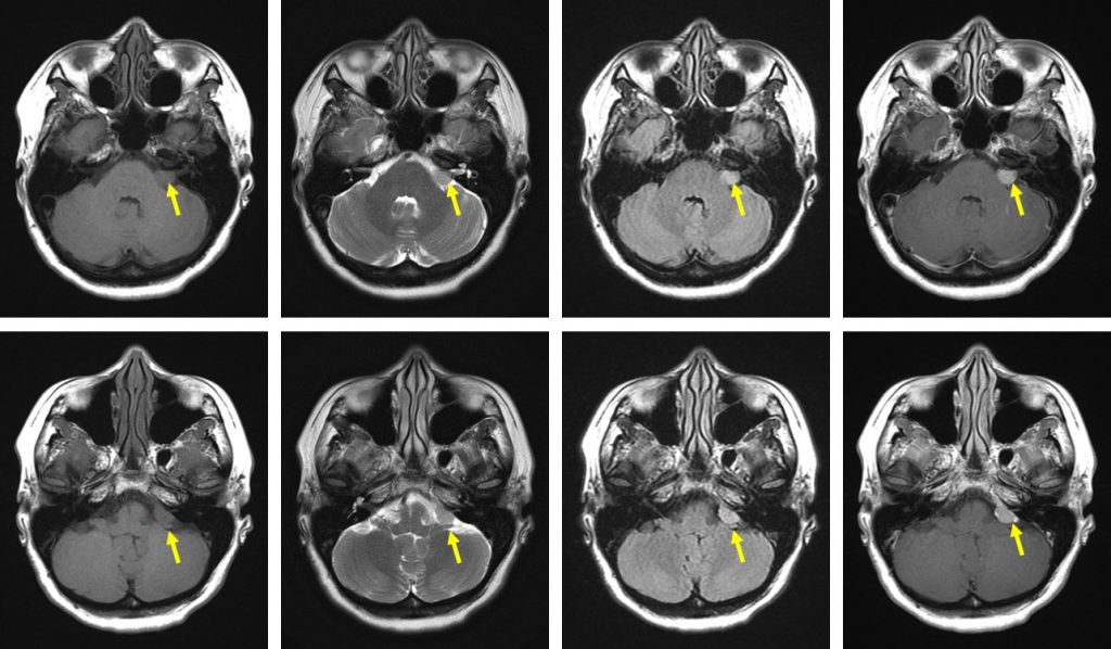
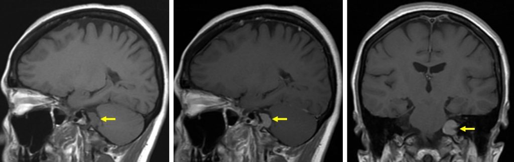
- There is an extra-axial mass lesion at left cerebello-pontine angle (Yellow arrows)
- This lesion is isointense on T1, hyperintense on T2, not suppressed on FLAIR and shows homogenous enhancement post contrast
- There is no blooming artefact on GRE and no restricted diffusion (images not shown)
- broad based attachment to the dura is also seen.
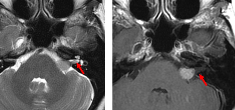
- Extension and causing widening at the opening of the adjacent internal auditory
- However the vestibulocochlear nerves are seen not enhanced (red arrows)
- No bone erosion or hyperostosis (ct images not shown)
Diagnosis: CPA meningioma
Discussion:
- CPA dural-based enhancing mass with dural tail sign (60% of cases)
- On T1, iso to minimally hyperintense to gray matter
- On T2Wi wide range of possible signal
- Calcification may bloom on GRE
- 95% enhances strongly, heterogenous enhancement if large lesion
- when extending into IAC may mimic vestibular schwannoma as in this case
Recent Comments