Case contribution: Dr Radhiana Hassan
Clinical:
- A 53 years with rectosigmoid adenocarcinoma
- Had sigmoid colectomy and left ureterotomy with stenting done
- Patient complaint of left sided abdominal pain on day 8 post operation
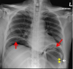
Chest radiograph findings:
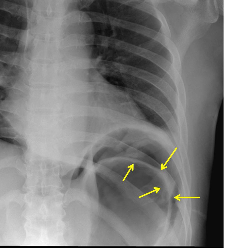
- Presence of free intraperitoneal seen
- Bilateral subdiaphragmatic free air (red arrows)
- Rigler sign (yellow arrow) with visualization of gastric wall
- Central venous line in-situ
- No pneumothorax
Radiological diagnosis: Pneumoperitoneum( subdiaphragmatic free air and Rigler sign)
Discussion:
- Free intraperitoneal gas can be seen following surgical procedures.
- It occurs in up to 60% of laparotomies and 25% of laparoscopic procedures.
- The volume of gas on serial radiographs should decrease.
- If volume is increasing, bowel perforation or anastomotic leak should be suspected.
- Signs of pneumoperitoneum on plain radiograph include subdiaphragmatic free air and rigler sign as seen in this case.
- Rigler sign is also known as double-wall sign of pneumoperitoneum seen when gas is outlining both sides of bowel wall. It is usually seen when there is large amounts of pneumoperitoneum (>1000mL)
- Other radiographic signs include cupola sign
Progress of patient:
- Subsequent imaging confirmed anastomotic leak in this case
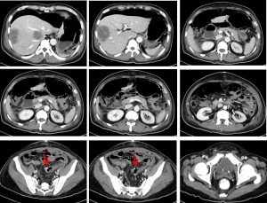
CT scan findings:
- A suspicious small discontinuity at rectal wall is noted (red arrows).
- Presence of loose surgical material at this region is also observed.
- Fluid is seen tracking from gap into the larger left paracolic gutter collection.
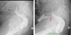
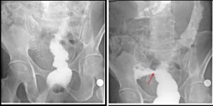
Lower GI contrast study done:
- Diluted Gastrografin (1:1) is infused via rectum through a Foley’s catheter 18F size, already anchored within the anal canal.
- Free cephalic flow of contrast is observed through rectum till the descending colon.
- Contrast extravasation from the anterior part of the rectum is observed approximately 12 cm from the anal verge, (red arrows) at the region of the anastomosis.
Recent Comments