Author: radhianahassan

Quiz 10

Image: Questions: Name the examinations. Name the probe used. Name the branches of ‘G’. Name the labelled stuctures. Answers: Ultrasound of upper abdomen in axial and sagittal plane. Curvilinear probe,…

 Posted in Quiz

Quiz 8

Image 1: Question: Name the labelled structures. What causes non-visualization or poor outline of “D”? Answers: The labelled structures A: right 12th rib B: fecal material/air in the ascending colon…

 Posted in Gastrointestinal Tract

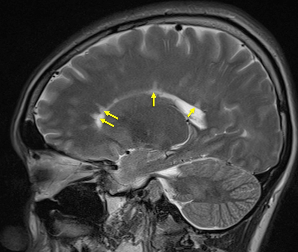
Dawson fingers (multiple sclerosis)

Clinical: A 22 years old lady Sudden onset of left sided weakness for 2 weeks Left sided loss of sensation since 13 days. Clinically had loss of left nasolabial fold…

 Posted in Central Nervous System

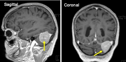
Tentorial meningioma

Clinical: A 79 years old lady Presented with left-sided blurring of vision for one month Associated with headache and dizziness. Clinical examination shows both optic discs are pale. No neurological…

 Posted in Central Nervous System, Tumour

Quiz 7

Image 1: Questions: Name the examination. Name the branches for ‘g’. Name the labelled stuctures. Answers: CT scan of abdomen, axial plane, soft tissue window, axial plane. Branches for g:…

 Posted in Quiz

Quiz 6

Image 1: Questions: Name the examination. Name the labelled structures. Give normal measurement of ‘i’. Answers: Radiograph of cervical spine in lateral view. The labelled structures: Mastoid air cells Anterior…

 Posted in Quiz

Quiz 5

Image 1 Questions: Name the examination. Name the labelled structures: What is pulmonary hilum and hilar point? What are possible disease process with an abnormal hilum? Answers: Chest radiograph, PA…

 Posted in Quiz

Quiz 4

Image 1 Questions: Name the examination. Name the labelled structures. Answers: MRI of neck in axial plane, T2-weighted image. The labelled structures: Right maxillary sinus Right masseter muscle Right lateral…

 Posted in Quiz

Quiz 3

Image 1 Questions: Name the ultrasound artefact shown. Briefly describe why it occurs. State how to rectify this. Answers: Reverberation artefact. Reverberation artefact occurs when ultrasound beam encounter strong parallel…

 Posted in Quiz

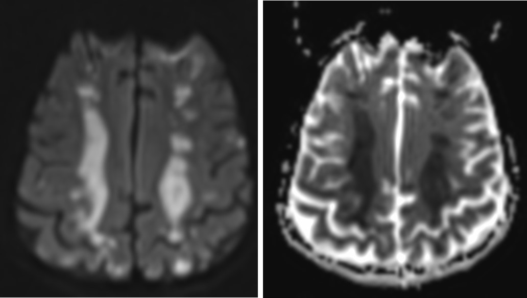
Watershed infarction

Clinical: A 16 years old boy Underlying liver failure Presented with poor GCS Also had one episode of hypotensive and bradycardia MRI findings: Areas of restricted diffusion in white matter…
 Posted in Central Nervous System