
Case contribution: Dr Radhiana Hassan
Clinical:
- A 55 years old man
- Involved in MVA
- Complaint of headache, GCS=14/15
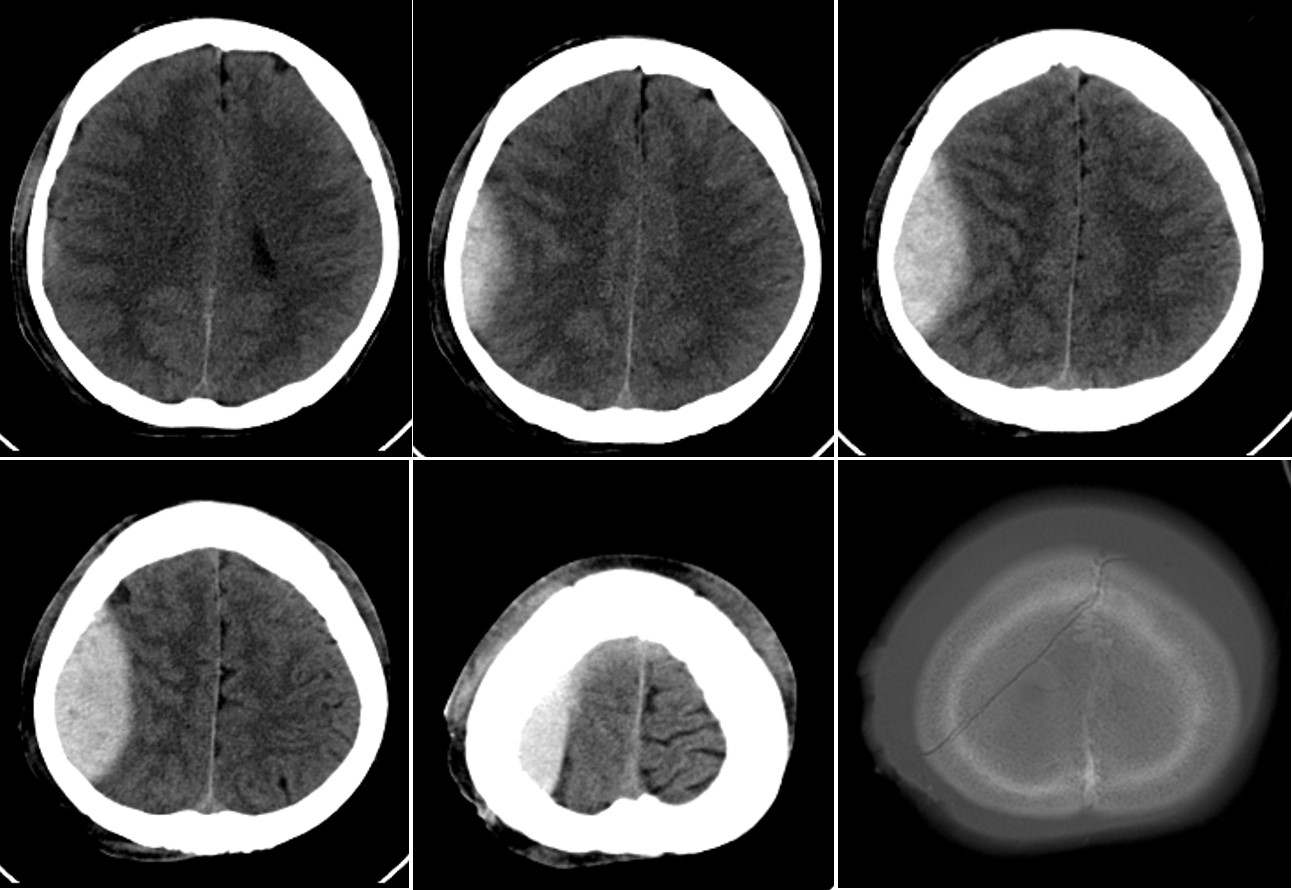
CT scan findings:
- A lens-shaped hemorrhage in the right fronto-occipital region
- It do not cross the suture
- Compression effect to the brain parenchyma
- Effacement of ipsilateral cerebral sulci
- No significant midline shift
- On bone window, a skull fracture is seen
Diagnosis: Traumatic right extradural hemorrhage
Discussion:
- also known as an epidural hemorrhage
- A collection of blood that forms between the inner surface of the skull and the endosteal layer.
- They are usually associated with a history of head trauma and frequently associated skull fracture.
- The source of bleeding is usually arterial, most commonly from a torn middle meningeal artery
- EDHs are generally unilateral in more than 95% of cases, however, bilateral or multiple EDHs are reported.
- Supratentorial location: temporoparietal (60%), frontal (20%) and parieto-occipital (20%)
- infratentorial location (5%) in posterior fossa
- Management is craniotomy with evacuation of blood
- Surgical intervention if
- EDH larger than 30 cm3, regardless of GCS
- EDH with GCS <9
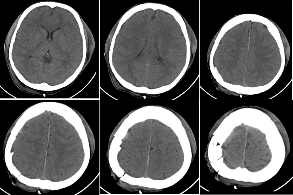
Progress of patient:
- Craniotomy and evacuation done
Recent Comments