
Clinical:
- A 49 years old lady
- Presented with progressive abdominal distension over one year
- No constitutional symptoms
- No bowel related symptoms
- Menstrual cycle is regular and normal
CT findings:
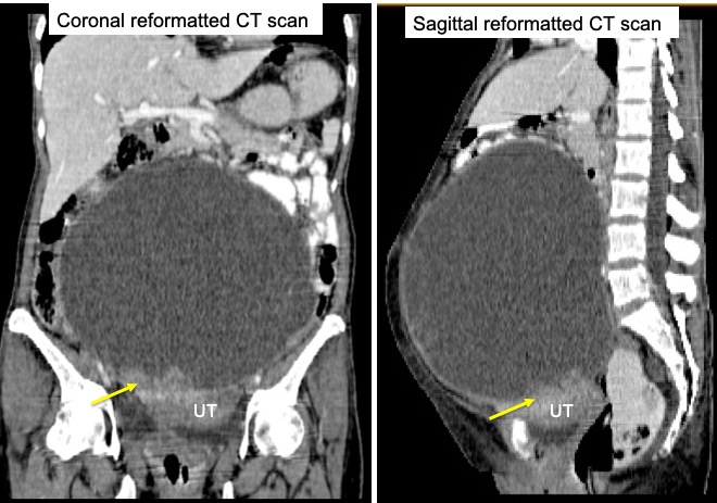
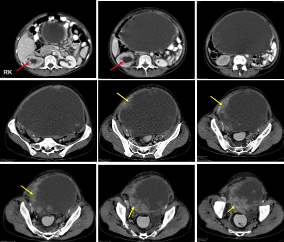
- A large cystic lesion arising from the pelvic region extending to upper abdomen
- The lesion is mainly cystic with no calcification or fat component within
- There are soft tissue components noted at its inferior part (yellow arrows)
- The soft tissue component showed enhancement after contrast
- No clear plane of demarcation with the uterus
- Right hydronephrosis also noted (red arrows)
- No ascites. Other organ are grossly normal
Intra-operative findings:
- A large right ovarian tumour 34x30x30 cm. Adherent to visceral peritoneum, small and large bowel. Ruptured during manipulation, contains blood stained fluid about 4 litres.
- Several enlarged external iliac nodes
- Left ovary small and normal looking
HPE findings:
- Macroscopy: specimen consists of uterus, cervix, left ovary, ruptured right ovarian cyst, both fallopian tubes, tumour nodule and multiple pieces of omentum. Cut open of right ovarian cyst shows wall thickness of 1mm to 15 mm. There are multiple nodules ranging from 18 mm to 30 mm in diameter. Cut section of the nodules show greyish cut surface.
- Microscopy: sections from the right ovary show a malignant tumour with extensive area of tumour necrosis. The tumour is composed of malignant cells arranged in solid sheets and tubopapillary structures infiltrating a sclerotic stroma. In solid area the tumour composed of predominantly tumour cells exhibiting round to oval vesicular nuclei with prominent nucleolus and abundant clear cytoplasm. However the tubopapillary structures in areas lined by tumour cells with high grade nuclear features displaying pleomorphic vesicular nuclei with large central nucleoli and moderate amount of cytoplasm. Many mitotic figures are seen in this area. Tumour deposits seen at serosal surface of uterus, right parametrium and omentum.
- Interpretation: Right ovary: clear cell adenocarcinoma
Diagnosis: Ovarian clear cell adenocarcinoma.
Discussion:
- Ovarian clear cell adenocarcinoma is a malignant epithelial tumour
- They represent about 2-5% of all ovarian carcinomas and 4-12% of epithelial ovarian neoplasms.
- Mean age of presentation is younger than other epithelial tumour with peak age at 55 years old.
- Macroscopically comprise of a large unilocular cystic mass with protruding solid nodules.
- On imaging, typically seen as unilocular mainly cystic smooth marginated mass with lumen protruding solid portion and high attenuated cystic portion.
- MRI signal varies often dependant on hemorrhagic component
Recent Comments