
Clinical:
- A 68 years old man
- Presented with chronic cough for about 6 months
- Recently also noted pain on deep inspiration
- Associated loss of appetite and loss of weight
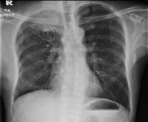
Chest radiograph PA erect view 
Radiographic findings:
- Hyperinflated lung fields bilaterally.
- Arounded opacity seen at right upper zone (yellow arrow). No cavitation or calcification within the opacity. No air bronchogram sign.
- There is associated erosion of posterior 3rd and 4th ribs (red arrows).
- No other lesion elsewhere. No mediastinal widening or shift
- No obvious hilar mass. No pleural effusion or pneumothorax.
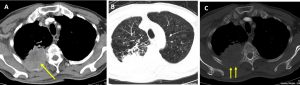
CT scan findings:
- There is a lobulated mass (yellow arrow) seen in the posterior segment of right upper lobe measuring 66 mm. The mass showed soft tissue density with heterogenous enhancement post contrast. No calcification or cavitation seen.
- Surrounding the mass lesion is extensive bronchiectasis and fibrosis (lung window).
- There is lytic destruction of adjacent right posterior 3rd-5th ribs (yellow arrows).
- There is no other mass or lung nodule.
- Generalised and extensive centrilobular emphysematous change of both lungs, predominantly in upper lobes (lung window).
- There is no pleural thickening or pleural effusion. No pericardial effusion.
- A few enlarged mediastinal nodes are seen.
HPE findings:
- Macroscopy: specimen labelled as tru-cut biopsy of lung mass, consist of multiple pieces of whitish tissue measuring 2 to 15 mm in length.
- Microscopy: section shows multiple strips of fibrous tissue harbouring malignant tumour arranged as irregular glands accompanied by area of necrosis. These malignant cells are lined by moderately pleomorhic tumour cells displaying nuclear pleomorphism, vesicular to hyperchromatic nuclei and prominent nucleoli. Abnormal mitotic figure is identified, intraluminal mucin is also noted. There is no squamous differentiation seen. In areas, skeletal muscle bundles are also noted.
- Interpretation: Adenocarcinoma
Diagnosis: Adenocarcinoma of lung.
Discussion:
- Lung adenocarcinoma is the most common histologic type of lung cancer, it represents 31% of all lung cancers
- It is under non-small cell carcinomas of the lung
- It is a malignant tumour with glandular differentiation or mucin production expression in different patterns and degrees of differentiation
- Adenocarcinomas are typically peripherally located and measure <4 cm in diameter; only 4% show cavitation.
- Hila or hila and mediastinal involvement is seen in 51% of cases on chest radiography
- One study stated two characteristic appearances on CT: either a localized ground glass opacity which grows slowly (doubling time >1 yr) or a solid mass which grows more rapidly (doubling time <1 yr)
- However, it is often not possible to distinguish between other histological types of lung cancer based on imaging findings.
- The classification of lung adenocarcinoma was revised in 2011 at which time the term bronchoalveolar carcinoma was removed.
- Four new terms were introduced to replace this term:
- adenocarcinoma in situ (AIS),
- minimally invasive adenocarcinoma (MIA),
- invasive mucinous adenocarcinoma and
- lepidic predominant non‐mucinous adenocarcinoma.
Recent Comments