
Case contribution: Prof. Dr Azian Abd Aziz
Clinical:
- A 9 year old girl
- Recurrent afebrile seizures.
MRI findings:
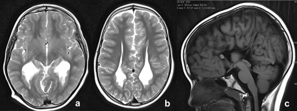
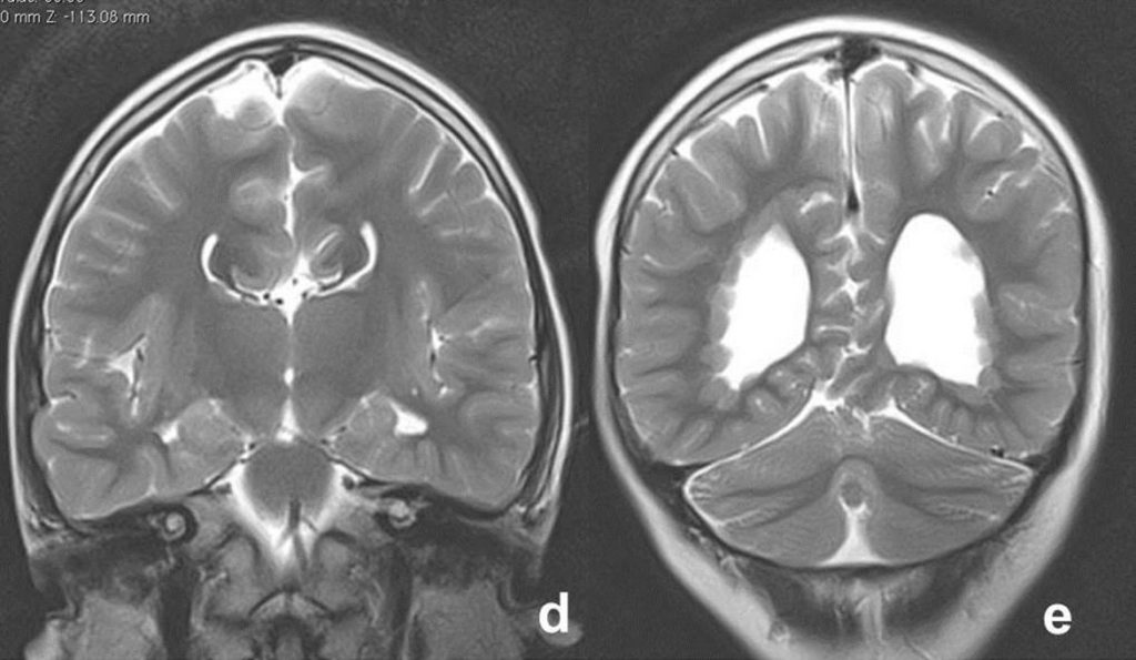
- MRI axial T2 (Figs. a & b), sagittal T1 (Fig. c) and coronal T2 (Figs. d & e).
- Body of the lateral ventricles is parallel to each other (Fig. b), absent of the cingulate sulcus with the medial hemispheric sulci reaching the third ventricle in a radial fashion (Fig. c), and a bicornuate or ‘Moose head’ appearance is displayed on the coronal view (Fig d) consistent with absent of the corpus callosum.
- Multiple subependymal nodules with similar signal intensity to the grey matter are seen at both occipital horns (Figs. a, b, e).
- Colpocephaly is observed bilaterally.
Diagnosis: Complete agenesis of corpus callosum with subependymal grey matter heterotopias and colpocephaly.
Discussion:
- Agenesis of corpus callosum may be complete, partial, or atypical.
- With complete agenesis, the corpus callosum is totally absent.
- Other cerebral malformations may coexist with callosal dysgenesis. Examples of these include interhemispheric cysts; intracranial lipomas; and disorders of neuronal migration, such as neuronal heterotopias, lissencephaly, pachygyria, and schizencephaly.
- Magnetic resonance imaging (MRI) is currently the imaging procedure of choice in infants and children with agenesis of corpus callosum, even in patients who have previously undergone CT and US examinations. The multiplanar capability and high soft-tissue contrast with MRI allows clear depiction of this condition and its associated intracranial anomalies. These appearances may not be well depicted on CT or US.