
Case contribution: Prof. Dr. Azian Abd Aziz
Clinical:
- A 1 year old boy
- Previously delivered prematurely at 33 weeks gestation and was followed up for hypoxic ischemic encephalopathy.
- During follow up, microcephaly and delayed development were noted.
- MRI was performed for further assessment.
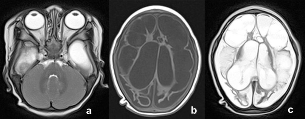
MRI findings:
- Multiple well defined cystic spaces similar to CSF on all sequences in both cerebral hemispheres involving both grey and white matter but sparing the brainstem and cerebellum.
- Asymmetrical ex-vacuo dilatation of the lateral ventricles is seen.
- Bilateral subdural effusion is also observed.
- The corpus callosum (not shown in the images) is thinned.
Diagnosis: Multicystic encephalomalacia
Discussion:
- Multicystic encephalomalacia or sometimes also known as subcortical leucomalacia is a condition where there are presences of necrotic areas that develop into cystic lesions in the brain
- It can be located in the cortical or white matter region.
- It is commonly seen in term neonates as a result of extensive brain insult from asphyxia due to hypoxic-ischemic injury. Other causes like meningoencephalitis and twin-to-twin transfusion may also result in this condition.
- The imaging appearance is usually associated with poor outcome.
- Cranial ultrasound (US) has been described as the primary investigative modality as compared to CT. This is well recognized as US is portable making it very convenient for bedside use especially in ill infants, can be quickly performed and no radiation is involved. US can also detect intracranial bleed if present and will also show ventriculomegaly, making necessary treatment be carried out promptly.
- MRI on the other hand demonstrates cavitations in the brain clearly especially with fluid attenuated inversion recovery (FLAIR) images where the cavities or cystic spaces are hypointense in the background of gliosis of the adjacent injured brain tissue.
- Although it may not be suitable for very sick infant, due to long examination time and monitoring issues, MRI findings of this condition can have great prognostic value which allows appropriate therapeutic options and adequate counselling to be provided to parents and caregivers.
Recent Comments