
Clinical:
- A 59 years old lady
- Fall on outstretched left hand
- Presented with pain at left wrist
- Clinically deformed swelling of left wrist
- “Piano sign” positive
- Radial pulse palpable
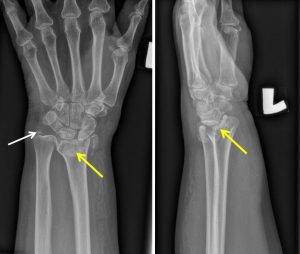
Radiographic findings:
- There is comminuted fracture of distal radius (yellow arrows)
- The fracture line involving the articular surface
- There is dorsal displacement of dorsal rim and carpus
- Fracture of ulnar styloid also seen (white arrow)
- Distal radioulnar joint is preserved
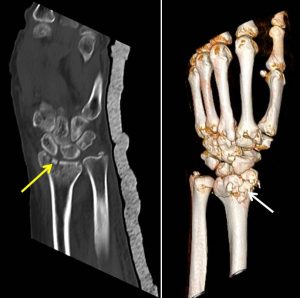
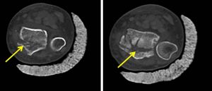
CT scan findings:
- There is a comminuted intra-articular fractures of the distal radius
- The fractures mainly involve the posterior half of the distal radius.
- Minimally displaced fracture fragments and intraarticular loose bodies are also seen, especially the radial aspect of the fracture.
- Minimally displaced ulnar styloid fracture is also seen.
- The radiocarpal joint is somewhat preserved.
- No angulation of the distal part of radius is detected.
- The Gilula’s arcs are preserved. No carpal bone fracture is detected.
Diagnosis: Dorsal type Barton fracture
Discussion:
- Barton fracture is fracture of distal radius
- In this case a dorsal type Barton fracture
- The fracture line extends through the dorsal aspect of the articular surface but not to volar aspect
- There is usually associated with dorsal subluxation or dislocation of radiocarpal joint
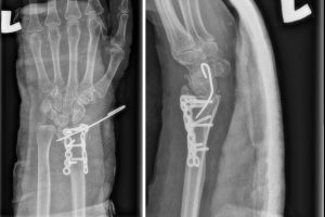
Progress of patient:
- Open reduction and locked plating done
Recent Comments