
Case contribution: Dr. Raja Rizal Azman
Clinical:
- A 10-year old girl presents with short stature secondary to Growth Hormone deficiency.
- She was born at term with no complications during delivery.
- She achieved all her developmental milestones normally.
- Apart from short stature there were no physical abnormalities on physical examination.
MRI findings:
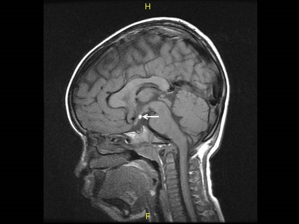
- T1-weighted sagittal image showing a focal hyperintense lesion at the floor of the third ventricle (white arrow).
- The normal bright stripe of the posterior pituitary in the posterior portion of the sella is absent.
- There is platybasia of the skull base.
- The residual pituitary gland within the sella turcica appears hypoplastic.
- The corpus callosum is preserved.
Diagnosis: Ectopic posterior pituitary with platybasia
Discussion:
- Ectopic posterior pituitary is a rare condition usually presenting with short stature.
- The location of the ectopic posterior pituitary is commonly at the median eminence at the base of the third ventricle.
- Ectopic posterior pituitary can be isolated or associated with numerous other conditions such as septo-optic dysplasia, platybasia, Chiari 1 malformation, corpus callosum dysgenesis, and Kallman syndrome.
- Careful evaluation of the MRI including thin slices of the midline and contrast enhanced images should be made to look for other abnormalities of the brain.
- The association with numerous other congenital midline abnormalities suggests a possible common underlying genetic aetiology.
Recent Comments