
Clinical:
- An 84 years old man.
- Admitted for acute urinary retention
- Subsequently diagnosed Benign prostatic hypertrophy
- Noted abnormal chest radiograph (incidental finding).
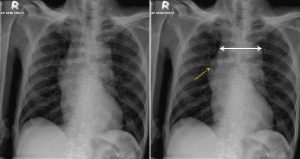
Radiographic findings:
- There is widening of mediastinum. The mediastinum measured at aortic arch level (double-head arrow) is 9.9 cm.
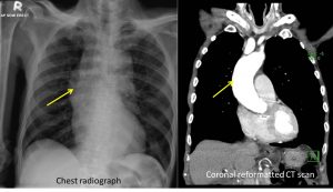
- There is a well-defined bulge at the right mediastinal outline (yellow arrow) which shows continuation with the aortic arch.
- No lung lesion seen. No pleural effusion or pneumothorax.
- Heart is not enlarged. No bone changes.
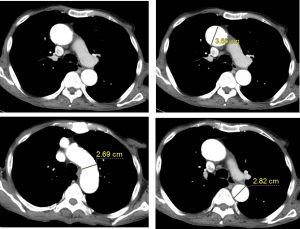
CT scan findings:
- The aortic root measures about 3.6 cm, while the aortic arch and descending aorta measuring about 2.7 cm and about 2.8 cm respectively.
- No aortic aneurysm or dissection is demonstrated.
- No intimal flap or thrombus seen.
- The heart is not enlarged. No pericardial effusion.
Diagnosis: Mediastinal widening caused by unfolded aorta.
Discussion:
- Unfolded aorta is not a pathological condition but can be mistaken for thoracic aneurysm.
- It is a common x-ray finding in elderly patient.
- It is seen as widened mediastinum on frontal chest radiograph. Normal mediastinal width should be <8cm at the level of aortic arch.
- Also descibed as “opened up” appearance of the aortic arch.
- This is due to discrepancy in the growth of ascending aorta with age
Recent Comments