
Clinical:
- A 56 years old lady
- No medical problem
- Presented with swelling at left plantar, non tender, slowly increased in size for the past few years.
- Clinically firm palpable plantar lumps felt
- The lesions were non-tender, no associated skin changes
MRI findings:
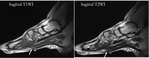
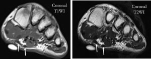
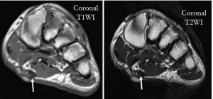
- Non-contrast-enhanced MR imaging of the left foot revealed nodular thickening along the medial band of the plantar fascia/aponeurosis (white arrows)
- The nodules are hypointense relative to muscle on the T1-weighted sequences and isointense to slightly hyperintense on T2-weighted sequences
- No infiltration to muscle or skin. The adjacent muscles and tendons are normal.
- There is no abnormal marrow signal of the visualized bones.
Diagnosis: Plantar fibromatosis.
Discussion:
- The plantar fascia, or aponeurosis, is a strong layer of fibrous tissue in the sole of the foot. It stabilises the medial longitudinal arch of the foot.
- Plantar fibromatosis, or Ledderhose disease, is a hyperproliferative disorder of mature fibroblasts that causes nodular thickening of the plantar aponeurosis.
- Although benign, it may invade the overlying skin or deeper structures such as the adjacent plantar muscles and tendon sheaths.
- Plantar fibromatosis can be seen in both children and adults. It occurs most frequently between the ages of 30 and 50 years
- There is a recognised male predilection (M:F of 2:1).
- Bilateral involvement seen in 20%–50% of cases.
- Concomitant palmar fibromatosis is seen in 10%–65% of patients.
- Nodules or masses of plantar fibromatosis are typically located in the middle to the medial aspect of the plantar arch.
- Lesions may be symptomatic because of a mass effect or invasion of adjacent muscles or neurovascular structures.
- In contrast to Dupuytren disease, flexion deformities usually do not occur.
- On ultrasound, it is often seen as a hypo to mixed echogenicity, discrete, fusiform, multinodular thickening of the plantar fascia located separately to the calcaneal insertion.
- The typical MR imaging appearance of plantar fibromatosis is a poorly defined, infiltrative mass occurring in the deep aponeurosis adjacent to the plantar muscles in the medial aspect of the foot. These lesions typically have heterogeneous signal intensity equal to or less than that of skeletal muscle on both T1- and T2-weighted spin-echo MR images. Lesions with high signal intensity on T2-weighted images reflect a more cellular lesion with relatively less collagen. The enhancement with gadolinium contrast material is variable, with marked enhancement seen in approximately 50% of lesions.
- Lesions with minimal symptoms are usually treated conservatively.
Recent Comments