
Clinical:
- A 63 years old lady
- Presented with vague fullness at lower abdomen region
- No altered bowel habit
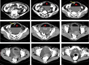
CT scan findings:
- There is a cystic mass measuring about 10x10x10 cm in the pelvic region
- It showed well defined outline with wall of moderate thickness and smooth in outline
- No enhancement post contrast
- Presence of calcification (yellow arrows) and fat component (star-shape)
- No ascites, no obvious infiltration to the surrounding structures
Intra-operative findings:
- There is left ovarian tumour 10×8 cm
- Solid cystic in nature, surface not smooth, upper part adhered to omentum, minimal vascularisation
- Whitish patches seen over surface of ovary
- Right ovary and fallopian tube are normal
- Uterus atrophic and normal
- No ascites
HPE findings:
- Macrosocpy: specimen labelled as TAHBSO consists of an atophic uterus, cervix, both fallopian tubes, right ovary and left ovarian mass. The left ovarian mass measures 110x90x100 mm. The capsules intact with smooth surface. Cut section shows uniloculated cyst filled with yellowish cheesy like material admixed with hairs. The cyst wall thickness ranging from 1 to 3 mm. No solid area seen.
- Microscopy: section from the left cyst wall show it is lined by a dingle layer of flattened epithelium and focally by foamy macrophages. In one area, the wall is lined by stratified squamous epithelium with keratin flakes. Within the cyst wall, there are skin appendages, such as sebaceous gland, hair follicles, fat cells, thyroid follicles, infarcted bone and cartilage and fibrous tissue. In other areas the wall is lined by respiratory type of epithelium. No immature tissue noted. The cervix is unremarkable. The uterus is atrophic. The right ovary shows focal calcification. Both fallopian tubes are normal.
- Interpretation: Benign cystic teratoma of the left ovary. No malignancy seen.
Diagnosis: Mature cystic ovarian teratoma.
Discussion:
- Mature cystic teratoma is the most common neoplasm of the ovary
- It is seen in a younger age group (mean age 30 years) than epithelial ovarian neoplasms.
- They are seen bilaterally in 12% of the cases . In unilateral cases, it occurs more frequently on the right side (72.2%) .
- Different combinations of mature tissue derivatives with varying arrangements in the tumour cause a wide spectrum of radiological findings ranging from a purely cystic mass to a complex cystic mass with a considerable solid component.
- Cross-sectional imaging methods (computed tomography and magnetic resonance imaging) have excellent sensitivity for the diagnosis due to identification of fat. In addition to detection of fat, other clues such as the fat-fluid level, floating ball sign, palm tree-like protrusion and reversed chemical shift artefact may be seen.
Recent Comments