
Clinical:
- An 18 years old girl
- Thallassemia major with regular blood transfusion
- Currently also had iron overload
Radiographic findings:
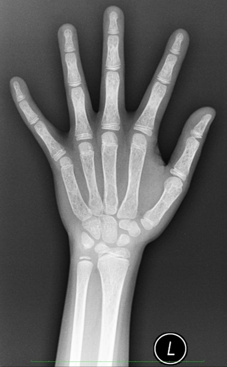
- There are coarse trabeculations involving the metacarpals, phalanges and carpal bones
- Expansion of these bones are also seen
- The cortices are thinned, however no fracture is seen
- No obvious deformity.
- Normal joint spaces
Diagnosis: skeletal changes in thalassemia
Discussion:
- The skeletal changes seen in thallasemia patients are due to marrow proliferation
- Within the medulla, first there is thinning of the trabeculae followed by their coarsening and expansion
- Thinning of cortical bone and resorption of cancellous bone resulting in a generalized loss of bone density and yellow-to-red bone marrow reconversion.
- The changes being most marked in the metacarpals and phalanges, which become cylindrical or biconvex in shape.
- Fractures may occur, although less commonly seen than expected from the degree of osteoporosis.
- Well-defined erosions of the periosteal margin of the cortex of the metaphysis or diaphysis may be identified in severe cases.
- Other bones commonly involved include the skull, the facial bones and the ribs.
Recent Comments