
Clinical:
- A 27 years old man
- Alleged MVA, motorbike skidded
- Complaints of painful swelling of right elbow
Radiographic findings:
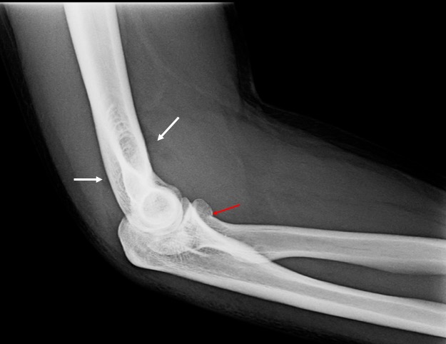
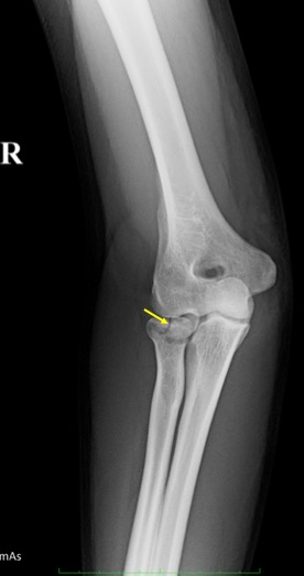
- A minimally displaced fracture is seen involving the radial head (red arrow)
- There is extension of fracture line to involve the articular surface (yellow arrow)
- Positive anterior and posterior fat pad signs are seen (white arrows)
- Associated soft tissue injury medial side of the elbow
Diagnosis: Fracture of radial head (Type II according to Mason classification)
Discussion:
- Radial head fracture is a common injury (half of adult elbow fractures)
- Most common cause of positive fat pad signs in adult
- Undisplaced fracture may be occult and requires several radiographic projections
- Mason classification of radial head fractures:
- type I: non-displaced radial head fractures (or small marginal fractures), also known as a “chisel” fracture
- type II: partial articular fractures with displacement (>2 mm)
- type III: comminuted fractures involving the entire radial head
- type IV: fracture of the radial head with dislocation of the elbow joint