
Clinical:
- An 8 years old boy
- Presented with fever and cough for one week
- Clinical examination shows crepitations at right lower zone
Radiographic findings:
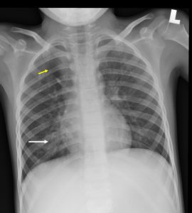
- There are air space opacities at right lower zone (white arrow). No obliteration of right cardiac margin or right hemidiaphragm is seen.
- Incidental finding of a curvilinear opacity (yellow arrow) at the right upper zone. This line shows smooth curve from the hilar superiorly towards the chest wall.
- No hilar mass, no pleural effusion or pneumothorax.
Diagnosis: Pneumonia with incidental finding of azygos fissure
Discussion (azygos fissure/lobe):
- It is found as an anatomic variant in about 1% of the population.
- The fissure contains both visceral and parietal pleural layers.
- On radiograph it is seen as a thin, curvilinear density in the upper right lung, convex towards the chest wall. At its base is the azygos vein which can be seen as a teardrop-shaped structure. The azygos vein will no longer be visible at its normal position at the junction of the trachea and right main bronchus
- An azygos lobe formed due to invagination of the azygos vein and pleura during development in the fetus. It has no bronchus so is not a true accessory lobe. The size of the lobe is variable
Recent Comments