
Clinical:
- A 13 years old girl
- Presented with suprapubic mass for 3 weeks
- There is associated pain which increased in severity
- No fever, no dysmenorrhea, menses are regular
- TAS shows a solid right adnexal mass
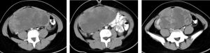
CT scan findings:
- There is a mass in abdominopelvic cavity measuring 17x15x8 cm.
- It is lobulated in outline with heterogenous density.
- Multiple septa within the mass lesion.
- The solid component and septa show enhancement post contrast.
- No calcification or fat component within it.
- Bowel loops are displaced peripherally.
Intra-operative findings:
- Right ovarian tumour 20×15 cm, lobulated surface, no breach of capsule. Contents mixed fleshy material with straw coloured cysts.
- Adhesion of mass with omentum, ruptured during manipulation.
- The left ovary and fallopian tubes are normal.
- Uterus is normal.
HPE findings:
- Macroscopy: Specimen labelled as right ovarian mass and fallopian tube. Serial cut section shows a variegated cut surface with greyish brown appearance with a few cystic spaces measuring 2 to 15 mm in diameter. There are also some haemorrhagic and mucoid cut surface.
- Microscopy: section show sheets of large neoplastic cells and at areas forming papillae. The cells exhibit marked nuclear pleomorphism and abundant eosinophilic cytoplasm. Mitotic activity is brisk. The cells are immunoreactive to alpha-fetoprotein, PLAP, vimentin, CD30 and beta HCG. There is extensive tumour necrosis.
- Interpretation: Germ cell tumour, features are in favour of embryonal carcinoma of right ovary with no evidence of metastasis to omentum. Metastic tumour in two pelvic nodes.
Diagnosis: Ovarian embryonal carcinoma
Discussion:
- Ovarian embryonal carcinoma is a rare malignant germ cell tumour of the ovary
- It is seen predominantly in children and adolescents (average 14 years old).
- The tumour can secrete beta HCG and/or AFP.
- On imaging it is predominantly solid, but nonspecific imaging features are seen that overlap with other germ cell tumours.
- Abnormal serum markers in the setting of an adolescent with a unilateral solid ovarian mass would put the tumour on a differential diagnosis.
Recent Comments