
Clinical:
- A 32 years old man
- Recently diagnosed retroviral positive with oncomycosis
- CT scan of thorax, abdomen and pelvis for assessment of his condition
CT scan findings:
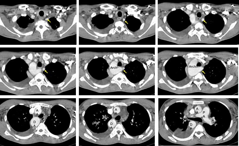
- Multisegmental collapsed of lungs due to proximal narrowing of its bronchi from external compression with multiple mediastinal and axillary lymphadenopathies.
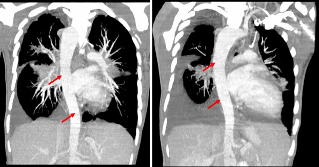
- There is right-sided aortic arch (red arrows).
- The left subclavian artery (yellow arrows) is seen coursing posterior to the esophagus before entering the aortic arch.
Radiological diagnosis: Incidental finding of right sided aortic arch with aberrant left subclavian artery.
Discussion:
- A right-sided aortic arch is thought to occur in approximately ~0.1% of the population.
- Right-sided aortic arch is a type of aortic arch variant characterized by the aortic arch coursing to the right of the trachea.
- Different configurations can be found based on the supra-aortic branching patterns
- Type I: right-sided aortic arch with mirror image branchin
- Type II: right-sided aortic arch with aberrant left subclavian artery (ALSA).
- Type III: right-sided aortic arch with isolation of the left subclavian artery
- The right sided aortic arch with aberrant left subclavian artery is common, accounting for 39.5% of all right-sided arches. It rarely produces symptoms and is usually an incidental finding. Rarely it may cause esophageal and/or tracheal compression. It is rarely associated with other cardiovascular abnormalities
Recent Comments