
Case contribution: Prof. Ahmad Sobri Muda
Clinical:
- An 80 years old lady. She is known hypertension on medication
- Presented with severe headache followed by loss of consciousness.
- She regain consciousness on arrival but GCS not full (13-14/15).
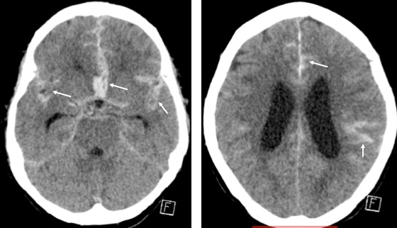
CT scan findings:
- There is extensive subarachnoid haemorrhage seen on CT without contrast (white arrows). Hunt and Hess at least grade II.
- The haemorrhages are most prominent at the interpeduncular cistern.
- No intraventricular haemorrhages or significant haemorrhage in the cerebellopontine cisterns.
- There is only minimal blood seen at the quadrigeminal cistern.
- Mild hydrocephalus is seen.
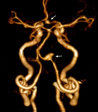
CT angiography findings:
- There are multiple aneurysms, with anterior communicating artery and left vertebral artery fusiform complex aneurysms (arrows).
- Irregular shape of both aneurysms but the SAH appears to be more prominent in interpeduncular cisterns, most likely the ruptured aneurysm is acom.
- Left vertebral artery aneurysm is most likely a dissecting aneurysm
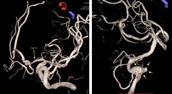
Cerebral angiogram images:
- Confirm presence of both vertebral and ACom Artery aneurysms
Diagnosis: Multiple brain aneurysms (anterior communicating and left vertebral) with most likely ruptured anterior communicating artery aneurysm.
Discussion:
- Aneurysms are focal abnormal dilatation of a blood vessel. They typically occur in arteries. Venous aneurysm are rare.
- Morphologically there are two main types of aneurysms.
- saccular aneurysm: eccentric, involving only a portion of the circumference of the vessel wall. (“Berry” aneurysm).
- fusiform aneurysm: concentric, involving full circumference of the vessel wall
- Prevalence of saccular cerebral aneurysms in the asymptomatic general population has been reported over a wide range (0.2-8.9%) when examined angiographically
- In 15-30% of these patients, multiple aneurysms are found.
- Cerebral aneurysms typically occur at branch points of larger vessels but can occur at the origin of small perforators which may not be seen on imaging. Approximately 90% of such aneurysms arise from the anterior circulation, and 15-30% of these patients have multiple aneurysms 4.
- anterior circulation: ~90%
- ACA/ACoA complex (30-40%), supraclinoid ICA and ICA/PCoA junction (30%) and MCA (M1/M2 junction) bi/trifurcation (20-30%)
- posterior circulation: ~10%
- basilar tip, SCA and PICA
- anterior circulation: ~90%
- Five-year cumulative risk of rupture of anterior circulation aneurysms:
- <7 mm: 0%
- 7-12 mm: 2.6%
- 13-24 mm: 14.5%
- >25 mm: 40%
Five-year cumulative risk of rupture of posterior circulation aneurysms:
- <7 mm: 2.5%
- 7-12 mm: 14.5%
- 13-24 mm: 18.4%
- >25 mm: 50%
Recent Comments