
Case contribution: Dr Radhiana Hassan
Clinical:
- A 32 years old lady
- Diagnosed fibrous dysplasia at 12 years old
- History of recurrent fracture involving left tibia and left femur since 10 years ago
- Since then having pain and left leg shortening
Radiographic findings:
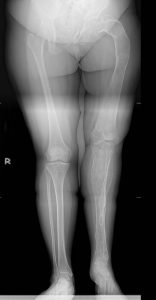
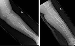
- There are homogeneous loss of trabecular pattern leading to ground glass appearance involving left femur, left tibia and left fibula.
- They show narrow zone of transition, with bony expansion.
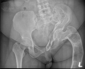
- Endosteal scalloping with cortical thinning is noted, with focal cortical defect at lateral aspect of left proximal femur. No callus formation is seen.
- Bowing deformity of the left femur is seen, with shepherd crook appearance of the femoral neck and bowing of the proximal femoral shaft laterally.
- No acetabulum protrusion is detected.
- There is shortening of left femur and left tibia and fibula.
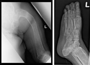
- Left hip, knee and ankle joints are unremarkable.
- No soft tissue mass.
Diagnosis: Fibrous dysplasia; polyostotic form (biopsy proven)
Discussion:
- It is a benign fibroosseous developmental anomaly of the mesenchymal precursor of bone, manifested as defect in osteoblastic differentiation and maturation
- Mean age of 8 years, about 2/3 are symptomatic by age of 10 years.
- Unilateral and asymmetric involvement; femur (91%), tibia (81%), pelvis (78%), foot (73%), rib, skull +facial bones (50%), upper extremities, lumbar spine (14%), clavicle (10%) and cervical spine (7%).
- Radiographic findings include leg length discrepancy, ‘shepherd’s crook’ deformity, tibial bowing, rib deformity and facial asymmetry.
- Malignant transformation seen in 0.5-1% of cases
- Common complication include pathological fractures.
Recent Comments