Case contribution: Dr Radhiana Hassan
Clinical:
- A 55 years old lady
- Presented with right loin pain
- Being investigated for urinary tract calculus and complex renal cyst

CT scan findings:
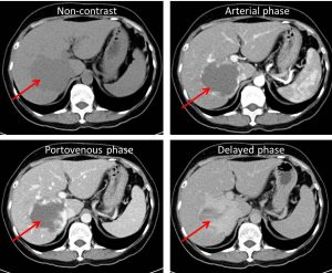
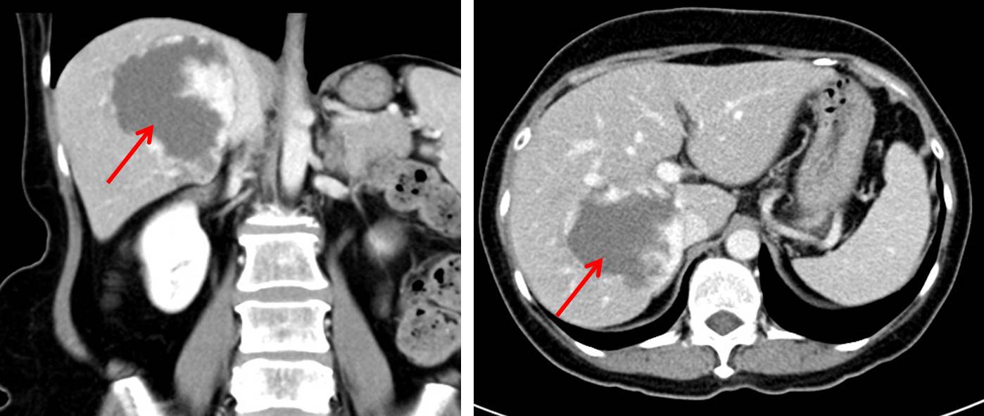
- A large well-defined hypodense lesion is seen occupying right lobe of liver measuring approximately 5.4 x 5.5 x 8.3 cm (AP x W x CC).
- No calcification of its wall.
- This lesion demonstrate discontinuous, nodular, peripheral enhancement on arterial phase.
- Progressive peripheral enhancement (centripetal fill-in) is seen on portovenous phase
- More irregular fill-in on delay phase making the lesion relatively hyperdense compared to the rest of the liver parenchyma.
Diagnosis: Hepatic venous malformation (hemangioma)
Discussion:
- Hepatic venous malformations also known as hepatic hemangiomas are the most common benign vascular lesion of the liver.
- It is more common in female with F:M ratio up to 5:1.
- It is considered congenital in origin.
- Ultrasound typically shows well-defined hyperechoic lesions.
- On CT scan, most hemangiomas are relatively well-defined. It is often hypodense on non-contrast study. On arterial phase it typically shows discontinuous, nodular, peripheral enhancement pattern, on portal venous phase it shows progressive peripheral enhancement with more centripetal fill-in. Delayed phase shows further irregular fill-in and therefore becomes iso or hyperdense compared to liver parenchyma.
- Complications are rare include spontaneous rupture, abscess formation and Kasabach-Meritt syndrome.
References:
- Hepatic hemangioma at https://radiopaedia.org/articles/hepatic-haemangioma
- Hepatic hemagioma; disorders of liver, biliary tract, pancreas and spleen. Wolfgang Dahnert.
