
Case contribution: Dr Radhiana Hassan
Clinical:
- A 28 years old lady
- Presented with amenorrhoea for one year
- No blurring of vision, no headache
- No constitutional symptom
- Investigation shows hyperprolactinaemia
MRI findings:
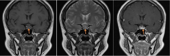
- Pituitary gland shows normal size and signal intensity.
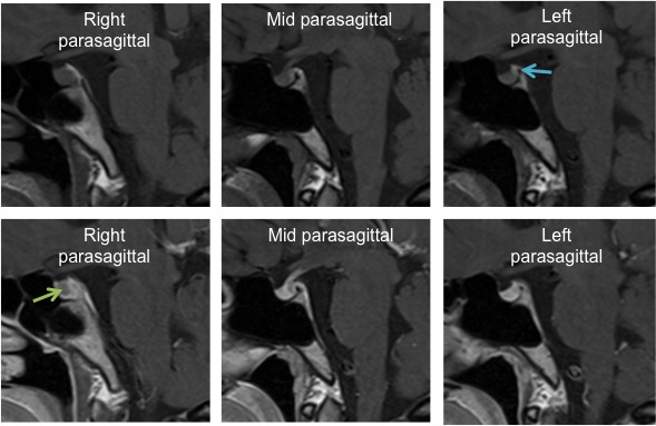
- Posterior lobe is identified at its normal location (blue arrow).
- However the right side of the lobe shows convex upper border (green arrow) compared to left side which maintains its superior concavity
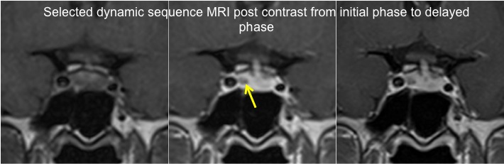
- Post contrast dynamic sequence shows a small relatively non-enhancing nodule in the right lobe measuring about 3 mm in diameter.
- The infundibulum is slightly deviated to the left side, otherwise normal in size and signal intensity
- No sella expansion
- No involvement of the optic chiasm
Diagnosis: Pituitary microadenoma
Discussion:
- MRI is the main imaging for pituitary microadenomas
- Dedicated pituitary sequences are important for diagnosis (thin slice, small field of view, dynamic contrast acquisition).
- Contrast-enhanced MRIs have a sensitivity of 90%.
- On T1 it is usually isointense to normal pituitary
- T1 dynamic sequences post contrast demonstrate a rounded region of delayed enhancement compared to the rest of the gland
- Appearance on delayed images are variable, ranging from hypo-enhancement (most common) to isointense to the rest of the gland, to hyperintense (retained contrast)
- On T2WI it is often variable, but often a little hyperintense
Progress of patient:
- Hormone level was reduced after medical treatment
- Patient also has regular menses
- Repeat MRI shows slight reduction in the size of the pituitary lesion.
Recent Comments