
Case contribution: Dr Radhiana Hassan
Clinical:
- A 66 years old man
- Type 2 DM on insulin
- Also had diabetic nephropathy and dyslipidaemia
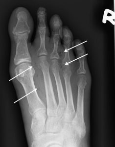
- Presented with diabetic foot
- Planned for amputation
- Pre-operative chest radiograph done
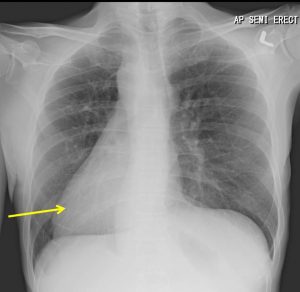
Radiographic findings:
- The heart shadow is seen projecting over the right side of the thoracic cavity
- The heart apex is pointing to the right side
- Otherwise the aortic arch is normally located
- No lung changes
- Left hemidiaphragm is higher on the left side
- Both costophrenic angles are sharp
- Air in gastric fundus is seen on the left side.
Diagnosis: Dextrocardia
Discussion:
- Dextrocardia is a congenital condition in which the heart is situated on the right side of the body with the apex pointing to the right.
- There are two main types of dextrocardia:
- dextrocardia of embryonic arrest (also known as isolated dextrocardia) and
- dextrocardia situs inversus (heart and visceral organs are mirrored on the right side)
- Dextrocardia is believed to occur in approximately 1 in 12,000 people 2.
Progress of patient:
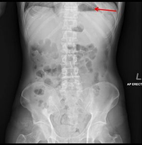
- Ultrasound abdomen shows normal position of liver and spleen
- No other abnormality of intra abdominal organ
- Echocardiography shows no abnormality of the heart
- Patient subsequently had BKA for diabetic foot with uneventful recovery
Recent Comments