
Case contribution: Dr Radhiana Hassan
Clinical:
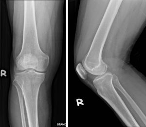
- A 71 years old man
- Underlying hypertension and IHD
- Complaint of right knee pain
- History of twisting knee with mild swelling
- Able to ambulate without aid
Radiographic findings:
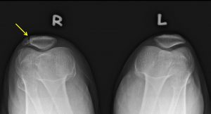
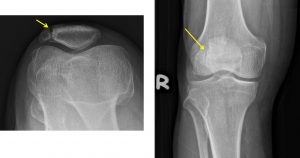
- There is a well-defined linear line seen at superolateral aspect of right patella (yellow arrows) separating the patella.
- The line has well corticated bone borders
- No displacement of the bone fragment
- No associated soft tissue swelling at knee region
Diagnosis: Bipartite patella
Discussion:
- A bipartite patella is a patella with an unfused accessory ossification centre at the superolateral aspect.
- Prevalence of bipartite patella is about 2% of population
- It occurs bilaterally in about 43% of cases.
- It is more common in male than females.
- Mostly are asymptomatic but patient can present with knee pain after trauma or overuse.
- Different location: superoplateral portion (75%), lateral margin (20-25%) and inferior pole (1%).
- Differentiation with fracture; most common patella fracture is transverse (linear fracture is very uncommon), fracture commonly occurs after direct trauma to knee and volume of fracture fragment usually equivalent of normal patella
Recent Comments