
Case contribution: Dr Radhiana Hassan
Clinical:
- A 16 years old boy
- History of fall during football game
- Complaint of left hip pain which was progressive associated with limping gait.
- On examination; flex left hip, abducted and externally rotated.
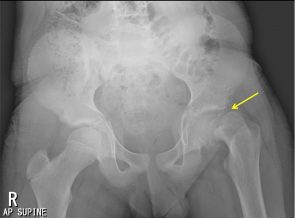
Radiographic findings:
- There is lateral displacement of the left proximal femur metaphysis, with widening and irregularity of left physis (yellow arrow).
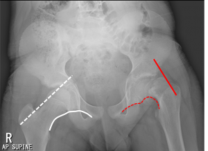
- Positive Threthowan sign is seen, in which the Klein line does not intersect with the left femoral epiphysis (red line). Compare to normal appearance on right side (white dotted line)
- There is also disruption of the Shenton’s line (dotted red line). Compare to normal line on the right side (white curve line).
- Positive metaphyseal blanch sign is noted, in which appearance of sclerosis of the metaphysis (due to overlap with the posteriorly displaced epiphyses).
- Left femoral epiphysis appear slightly smaller than the right side.
- The hip joint itself is otherwise normal.
- No flattening of the femoral head.
Diagnosis: Slipped upper femoral epiphysis
Discussion:
- Slipped upper femoral epiphysis is also called slipped capital femoral epiphysis.
- It is commonly affecting adolescents
- More common in boys than girls
- Obesity is a significant risk factor
- Other risk factors include hypothyroidism, hypopituitarism, hyperparathyroidism, renal osteodystrophy and radiation/chemotherapy
- Radiological findings include:
- The affected epiphysis appears smaller due to posterior slip
- Line of Klein fails to intersect the epiphysis (Trethowan sign)
- Loss of triangular sign of Capener ( metaphysis do not overlap with posterior lip of acetabulum)
- Metaphyseal blanch sign; increased density of proximal metaphysis due to superimposition of the femoral neck and posteriorly displaced capital epiphysis
- Treatment: surgical stabilization
- Complications:
- Osteoarthritis (90%)
- Avascular necrosis of the femoral head (10-15%)
- Chondrolysis (7-10%): acute cartilage loss
- Deformity-limb length discrepancy
- Femoroacetabular impingement
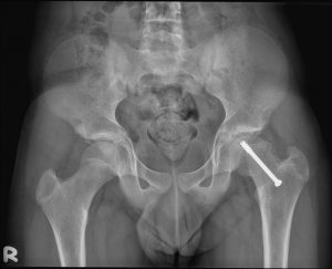
Progress of patient:
- Screw fixation done
- Uneventful recovery
- Able to play sport again
Recent Comments