
Case contribution: Dr Radhiana Hassan
Clinical:
- A 62 years old female
- Underlying DM and HPT
- Presented with worsening body weakness and facial asymmetry of few days
- Later had altered consciousness
- GCS at ED E3V5M5, BP=169/79mmHg and PR 69 bpm
- Power right upper and lower limb 4/5, left side 1/5
- One day after admission GCS E1V1M5
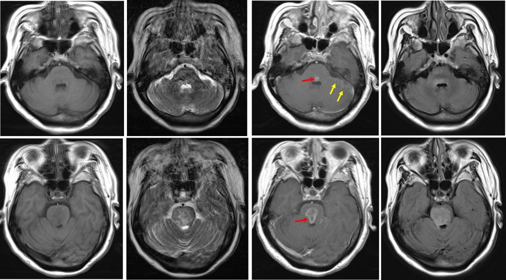
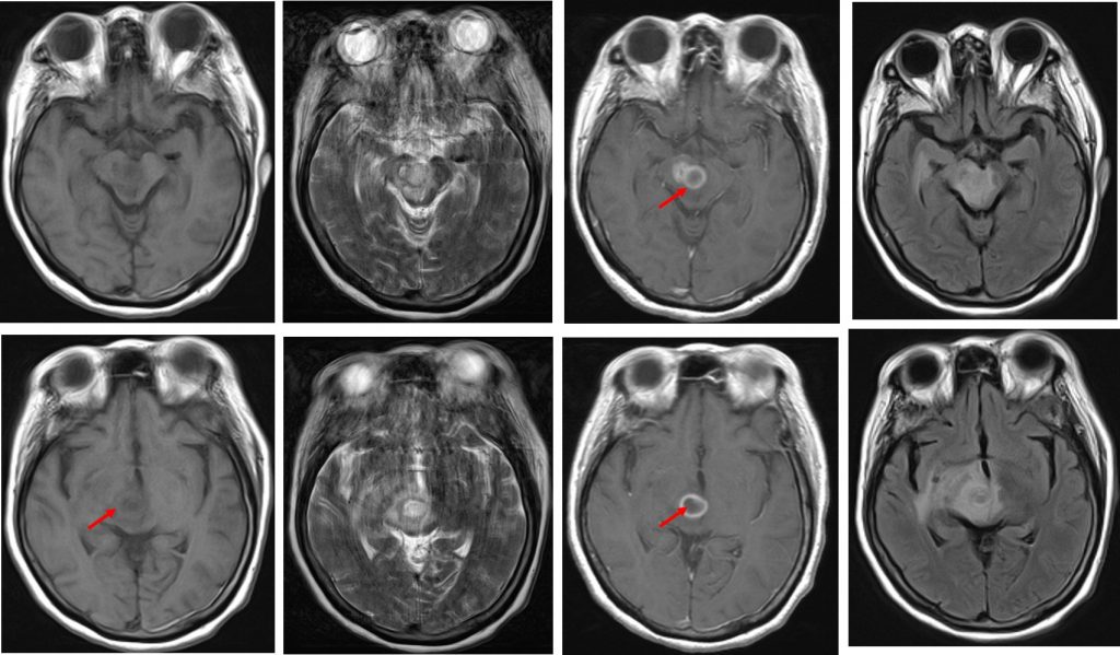
MRI findings:
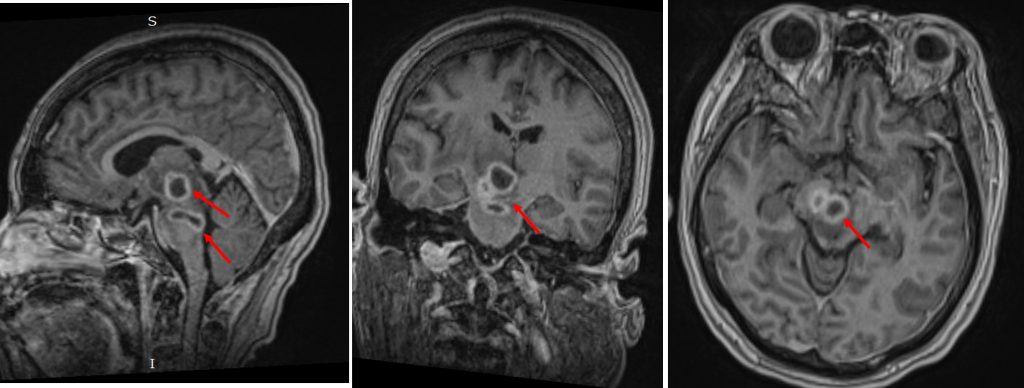
- There is ring-enhancing lesions in the right thalamus (red arrows)
- The wall is smooth with regular enhancement.
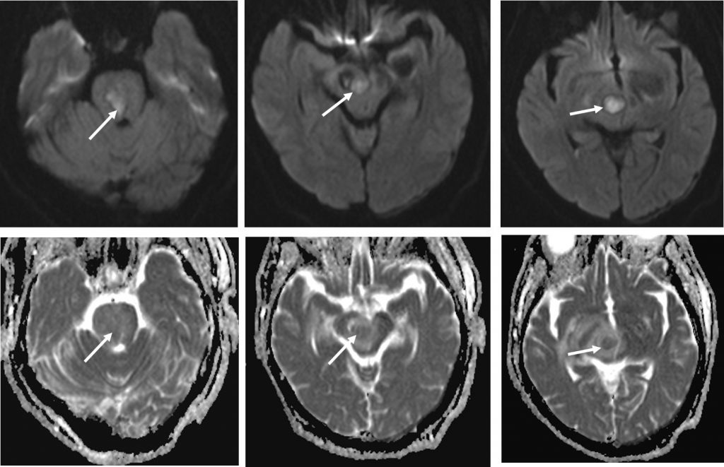
- Central part is hypointense on T1, hyperintense on T2/FLAIR and shows restricted diffusion on DWI/ADC sequences
- Smaller lesion seen adjacent to it with similar characteristic
- Another lesion with similar appearance but slightly irregular wall is seen at pons
- It is associated with significant surrounding oedema
- Leptomeningeal enhancement at left side of the cerebellar region (yellow arrows)
- No hydrocephalus, no basal cistern enhancing lesion
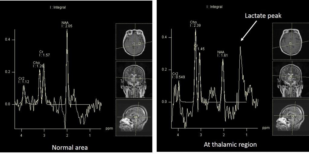
- MRS shows lactate peak
Diagnosis: Cerebral abscess
Discussion:
- Cerebral abscess represent focal areas of infection within brain parenchyma.
- It is usually pus-containing lesion with a thick capsule.
- They typically have enhancing walls and can mimic a number of other significant pathologies.
- It can occur at any age. Risk factors include immunocompromised (HIV & diabetes mellitus), existing infection (middle ear infection or bacteraemia) and IV drug use.
- CT and MRI demonstrate similar features.
- Typical appearance on CT include outer hypodense and inner hyperdense (double rims sign), ring of iso or hyperdense tissue of uniform thickness, central low attenuation region (pus/fluid), surrounding low density (vasogenic oedema), ventriculitis may be present. Obstructive hydrocephalus when intraventricular spread occured
- MRI is more sensitive; T1WI with central hypodensity and peripheral oedema, T2/FLAIR central hyperintensity , peripheral vasogenic oedema, DWI/ADC shows restricted diffusion, MRS shows elevated lipid/lactate succinate and acetate
Progress of patient:
- FBC: Hb 9.9, Plt 349 and TWBC 7.9 (all normal)
- RP: Urea 5.9, Na 130, K 4.4, Creat 85 (normal)
- LFT normal
- HIV rapid test negative
- CRP increased
- Sputum for TB negative
- CSF for TB no culture, no growth
- Patient responded well to antibiotics given, GCS 15/15
Recent Comments