
Case contribution: Dr Radhiana Hassan
Clinical:
- A 55- year- old man
- He had underlying diabetes mellitus , hypertension and valvular heart disease.
- Presented with shortness of breath and cough.
- There was no associated chest pain or fever.
- Clinically this patient had congestive cardiac failure, NYHA Class II.
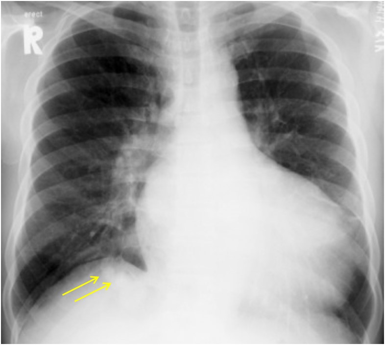
Chest radiograph findings:
- There are well-defined nodular opacities in the right lower zone below the right hemidiaphragm (yellow arrows)
- Patient also had gross cardiomegaly
- No feature of congestive cardiac failure. No pleural effusion.
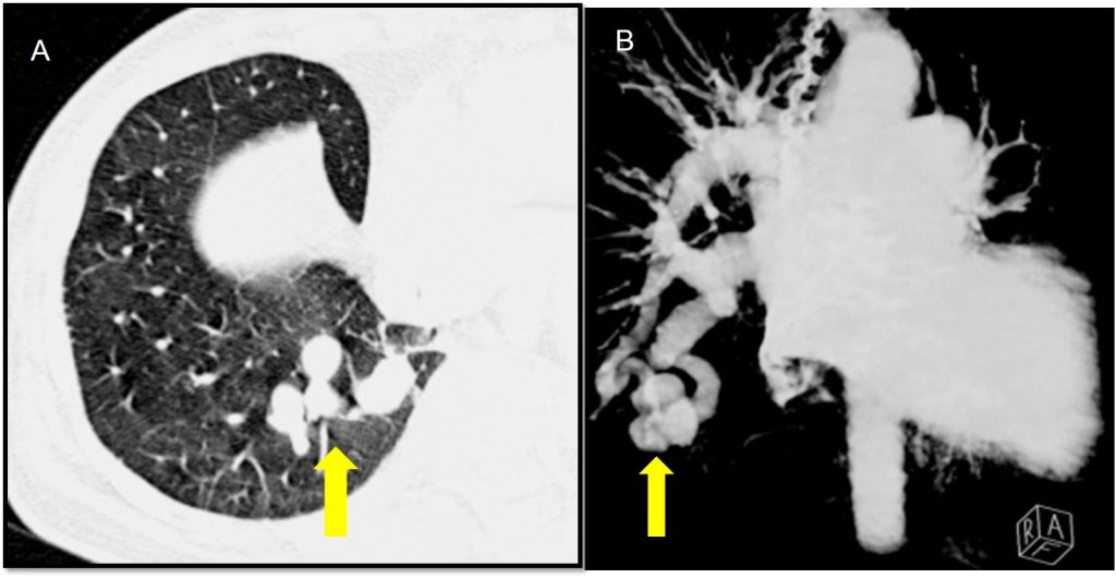
CT scan findings:
- Axial CECT image , lung window setting (A) shows multiple nodular lesions within the right lower lobe (block arrow)
- It shows similar density with vessels on post contrast images
- MIP image (B) shows the nodular densities were arteriovenous malformation with a single supplying artery and draining vein.
Diagnosis: Pulmonary arteriovenous malformation.
Discussion:
- Pulmonary arteriovenous malformation is not a common clinical problem.
- The estimated incidence is thought to be around 2-3 per 100,000 population.
- Mostly are asymptomatic, however patient can present with dyspnoea due to right-to-left shunting.
- It is often unilateral. Although can potentially affect any part of the lung, there is a recognised predilection towards the lower lobes (50-70%).
- Radiologically, this condition is an important differential diagnosis of pulmonary nodules detected on chest radiograph.
- CT is often the diagnostic imaging modality of choice. The characteristic presentation of a PAVM on non-contrast CT is a homogeneous, well-circumscribed, non-calcified nodule up to several centimetres in diameter or the presence of a serpiginous mass connected with blood vessels. Post contrast image demonstrates enhancement of the feeding artery, the aneurysmal part, and the draining vein on early-phase sequences.
- They can be classified as simple, complex or diffuse.
- simple type: commonest; has a single segmental artery feeding the malformation; the feeding segmental artery may have multiple subsegmental branches that feed the malformation but must have only one single segmental level
- complex type: have multiple segmental feeding arteries (~20%)
- diffuse type: rare (~5% of lesions); the diffuse form of the disease is characterised by hundreds of malformations; some patients can have a combination of simple and complex AVMs within a diffuse lesion
Reference:
Elfeky M et al, Pulmonary arteriovenous malformation at https://radiopaedia.org/articles/pulmonary-arteriovenous-malformation
Recent Comments