
Case contribution: Dr Radhiana Hassan
Clinical:
- A 44 years old lady
- No known medical illness
- Complained of right breast mass for the past 2 years
- Gradually increase in size and associated with occasional pain.
- No nipple discharge. No family history of breast cancer.
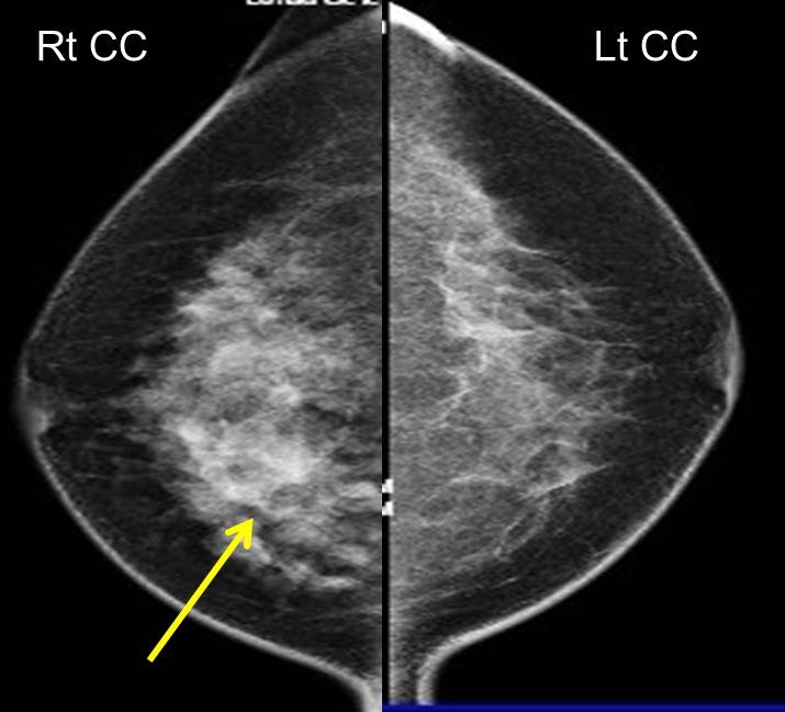
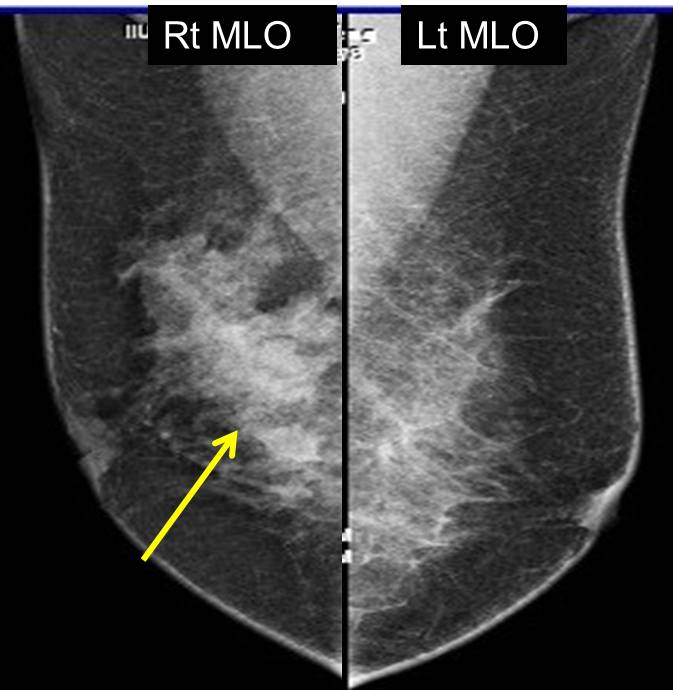
Mammogram findings:
- An area of increased density seen in the right breast (arrows)
- No obvious mass lesion is seen
- No clustered microcalcificaton
- No stromal distortion
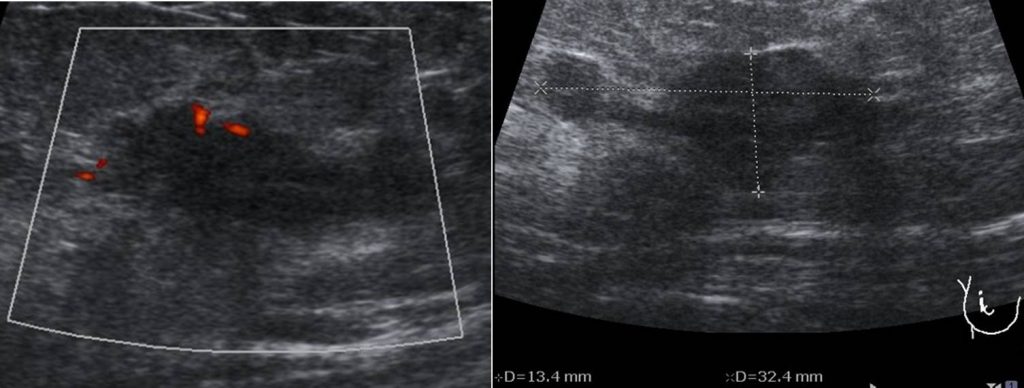
Ultrasound findings:
- A lobulated hypoechoic lesion is seen at upper quadrant of right breast
- Associated mild posterior shadowing
- It measures approximately 30 x 15 x 13 mm.
- Increased in vascularity within.
- Smaller lesion is seen adjacent to this lesion, with possible connecting duct
- No dilated duct is seen
Diagnosis: Benign intraductal papillary lesion (HPE proven)
Discussion:
- Intraductal papillomas are the most common masses within the milk ducts of the breast.
- They are benign tumors but may contain areas of atypia or carcinoma.
- Multiple intraductal papillomas arise from the terminal ductal lobular units and therefore are usually peripherally located in the breast.
- They are less common than solitary intraductal papillomas and rarely associated with a nipple discharge, and they typically present as a palpable mass.
- Unlike solitary papillomas, multiple intraductal papillomas are usually associated with atypia, DCIS, or malignancy. Some studies have shown that in patients with multiple papillomas, up to 80.4% may have either coexisting atypical lesions (atypical ductal hyperplasia (ADH), atypical lobular hyperplasia, lobular carcinoma in situ) or neoplastic lesions
- On mammography, small papillomas can be occult, particularly when located in the retroareolar regions. Larger lesions may appear as a round- or oval-shaped mass with well-circumscribed margins.
- On galactography, intraductal papillomas appear as well-defined mural-based filling defects with smooth or lobulated contours.
- On ultrasound, intraductal papillomas may appear as well-defined solid nodules or mural-based nodules within a dilated duct. On color Doppler imaging, flow may be detected within the papilloma arising from a vascular feeding pedicle.
Reference:
- Papillary lesions of the breast athttps://www.ajronline.org/doi/10.2214/AJR.11.7922
Recent Comments