
Case contribution: Dr Radhiana Hassan
Clinical:
- A 38 years old lady
- Presented with left breast lump for few weeks
- Mother had breast cancer at 6o years old
- Married and nulliparous
Mammogram findings:
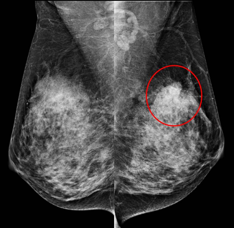
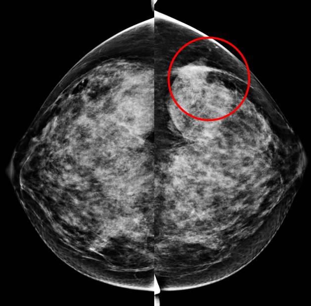
- Mammogram showed bilateral dense breasts, BIRADS C parenchymal density.
- A focal density seen at the left upper outer region.
- No obvious mass lesion is seen.
- No suspicious clustered microcalcification.
- No stromal distortion.
Ultrasound findings:
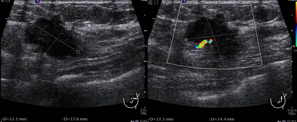
- A multilobulated lesion at Lt2H with irregular finger-like projection.
- It is measuring about 17x13x11 mm.
- There is associated posterior shadowing.
- Presence of penetrating vessels seen.
Progress of patient:
- Biopsy revealed invasive carcinoma
- Mastectomy done in another hospital
- Subsequently had chemotherapy
Discussion: Focal asymmetry on mammography
- A focal asymmetric densities are frequently encountered at screening and diagnostic mammography.
- These findings are significant because they may indicate a neoplasm, especially if an associated palpable mass is present.
- A focal asymmetric density is defined as density seen on two mammographic views but cannot be accurately identified as a true mass.
- They lack the convex borders of masses and are often interspersed with fat.
- They also lack the radiating lines or tissue retraction of architectural distortion (AD) and the tubular branching appearance of a dilated duct.
- Although a focal asymmetric density may represent normal breast tissue, further evaluation is often warranted to exclude a true mass or architectural distortion.
- To assess the shape and margins of a potential lesion, a spot compression view is obtained. If a density is clearly evident on two views but appears less dense or less prominent on the spot compression view, one should not assume that it is not a true lesion: Spot compression displaces the normal tissue away and may make a true lesion appear less dense
- US can also provide valuable information. The presence of a mass at US, particularly a hypoechoic solid mass or focal shadowing, raises suspicion for malignancy and definitely warrants biopsy. US can also demonstrate a cyst within a focal density that might prompt routine follow-up
- Causes of focal asymmetry include normal variation, post trauma, post surgery, sclerosing lobular hyperplasia, diabetic mastopathy and breast cancer
References:
- Focal asymmetric densities seen at mammography: US and pathologic correlation. RadioGraphics 2002 at https://doi.org/10.1148/radiographics.22.1.g02ja2219
- Mammographic asymmetries at radiologykey.com
- Asymmetry (mammography) at radiopedia.org
Recent Comments