
Case contribution: Dr Radhiana Hassan
Clinical:
- A 16 years old man
- Motorbike skidded and fall
- Complaint of pain at both upper limb
- Referred to our centre for further management of fractures
Radiographic findings:
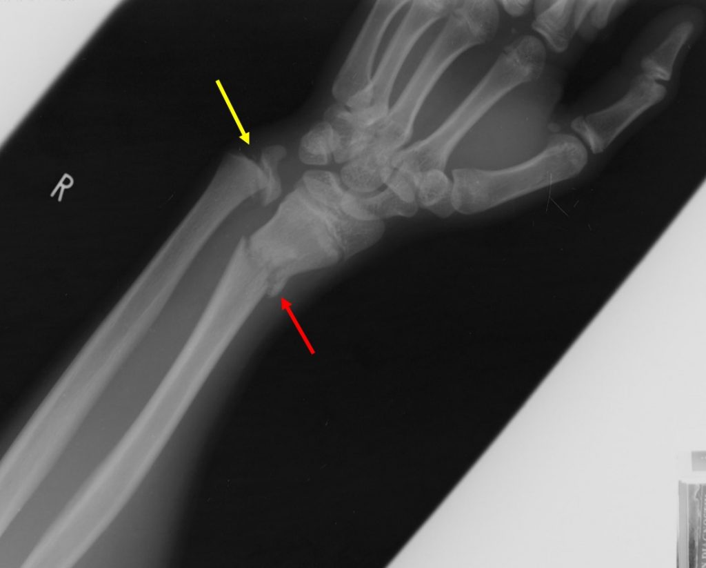
- There are horizontal fractures of both distal radius (red arrows)
- Displacement of fracture fragment on the left side
- Minimal angulation of right radius fracture fragment
- Separation and displacement of ulna physis
- No obvious fracture at metaphysis or epiphysis is seen
Diagnosis: Salter Harris fracture type 1 of distal ulna
Discussion:
- Salter-Harris fractures are fractures through a growth plate in pediatric patients.
- These fractures are categorized according to the involvement of the physis, metaphysis, and epiphysis.–Type 1: fracture plane passes through the growth plate and not involving the bone. Good prognosis. Seen in 5-7% of cases.
–Type II: fracture passes across the growth plate an through the metaphysis, the most common (75%) and also has good prognosis.
–Type III: fracture plane passes across most of the growth plate and down through the epiphysis, occurs about 7-10% of cases with poorer prognosis.
–Type IV: fracture plane passes directly through the metaphysis, growth plate an down the epiphysis, occur in about 10% of cases with poor prognosis.
–Type V: crushing type of injury which does not displace the growth plate but damages it by direct compression. Uncommon injury seen in less than 1% of cases. It carries worst prognosis.
- The classification of the injuries is important, because it affects patient treatment and provides clues to possible long-term complications.
-Grade I an II : Can be treated with close reduction an casting or splinting.
–Grade III and IV: Usually treated with open reduction an internal fixation.
Progress of patient:
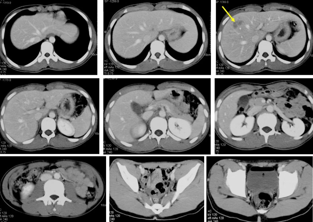
- During admission noted to have tenderness at abdomen on palpation
- BP=148/80mmHg, PR=70 bpm and remain stable
- CT scan shows grade II liver injury (yellow arrow)
- Parent decided took AOR discharged and refuse further medical treatment, opted for alternative treatment
Recent Comments