Case contribution: Dr Radhiana Hassan
Clinical:
- A 64 years old man
- Underlying HPT and CVA, gastric adenocarcinoma-lesser curvature
- CT scan abdomen as part of investigation for tumour.
- Patient has no urinary symptoms


CT scan findings:
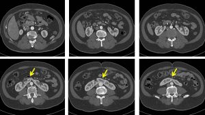
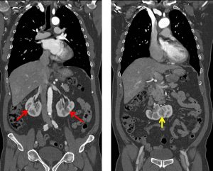
- Both kidneys are malrotated (red arrows)
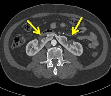
- The lower poles are fused together at midline (yellow arrows).
- No renal calculus or hydronephrosis.
- Urinary bladder is well distended and normal.
- Prostate is not enlarged, measures about 3.8cm x 3.8cm x 3.2cm (AP x W x CC).
Diagnosis: Horseshoe kidney
Discussion:
- Horseshoe kidneys are the most common type of renal fusion anomaly. It is usually asymptomatic and diagnosed incidentally.
- It is more common in males.
- It is formed by fusion across the midline of two distinct kidneys by functioning renal parenchyma or fibrous tissue.
- The kidneys are low lying and susceptible to trauma.
- There is also has higher risk for renal calculi and transitional cell carcinoma. Other complications include hydronephrosis, infection, pyeloureteritis cystica and renovascular hypertension.
- Horseshoe kidneys are frequently associated with genitourinary and non-genitourinary malformations.
- CT and MRI demonstrate renal tissue of normal imaging appearance but abnormal configuration. Enhancement and excretory phases are normal.
- Common features on imaging include:
- Midline symmetrical fusion of lower poles in 90% of cases or lateral asymmetric fusion in 10% of cases
- Position of fused kidneys are lower than normal with incomplete medial rotation
- Renal pelvis and ureters are positioned more anteriorly and ventrally cross the isthmus
- Isthmus that may be positioned below the inferior mesenteric artery
- Variant arterial supply that can originate from the abdominal aorta or common iliac arteries
- Lower poles of kidneys that extend ventromedially and may be poorly defined.
