
Case contribution: Dr Radhiana Hassan
Clinical:
- A 78 years old lady
- Had a total right hip replacement
- Post operative assessment complicated by DVT
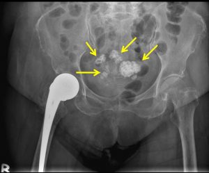
X-ray findings:
- Intact right hip prosthesis
- Incidental findings of multiple calcifications within pelvis
- No obvious mass lesion
- No dilated bowel loops
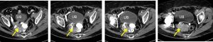
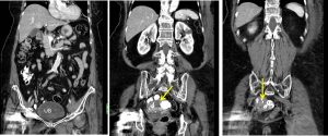
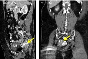
CT scan findings:
- Thrombosis of left external iliac and left femoral vein (images not shown)
- Uterus is bulky with multiple calcified lesions within its wall suggestive of calcified uterine fibroid (yellow arrows)
Diagnosis: Calcified uterine fibroid
Discussion:
- Uterine fibroid or leiomyomas are the commonest uterine neoplasms
- They are benign tumours of smooth muscle origin with varying amounts of fibrous connective tissue
- They are multiple in 85% of cases
- These tumour are hormone dependant and often increase in size during pregnancy and decrease in size after menopause.
- Calcification occurs in about 4% of fibroids and it is typically dense and amorphous. Calcification can also be found at the periphery of the fibroid and thought to be secondary to thrombosed veins from previous red degeneration
- Most calcified fibroids don’t require treatment.
Recent Comments