
Case contribution: Dr Radhiana Hassan
Clinical:
- A 53 years old lady
- No known medical illness
- Presented with right sided abdominal mass for 6 months
- Progressively increasing in size
- No constitutional symptoms, no altered bowel habit
- No jaundice, no vomiting
- Clinically abdomen is distended but soft, mass felt at epigastric extending to right lumbar, smooth surface and non-tender
- LFT normal, Hepatitis screening was negative, OGDS normal
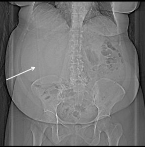
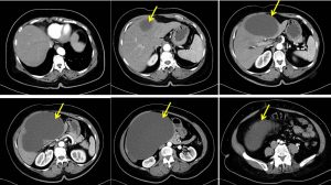
CT scan findings:
- Liver is normal in density with smooth outline and homogenous enhancement.
- There is a large cystic mass (HU 12) arising from segment III, IVb, V, and VI of the liver measuring 13.5 cm (AP) x 19.5 cm (W) x 13.9 cm (CC).
- This lesion has no internal septation or soft tissue component.
- Minimal wall calcification noted.
- No enhancement seen on arterial, venous and delay phase.
- The inferior vena cava (IVC) is partially compressed.
- No dilatation of biliary tree.
- The portal vein, coeliac trunk and its branches, and superior mesenteric artery are patent.
Operative findings:
- Laparoscopic deroofing and cyst aspiration of liver
- Intra-operative findings: upon entering peritoneum, there is huge cyst at surface of right lobe of liver . Aspiration shows straw coloured fluid with no pus or blood-stained content. Liver otherwise looks normal. Gallbladder is also normal.
HPE findings:
- Macroscopy: specimen labelled as liver capsule cyst, consists of multiple pieces of whitish tissue
- Microscopy: sections of the cyst wall show a well-defined fibrous capsule lined by flattened to simple columnar epithelium displaying uniform, basally-located nuclei, bland chromatin, inconspicuous nucleoli and abundant mucinous cytoplasm. No atypical cells or mitotic figures. Microcalcifications are noted within the cyst wall. In areas, the underlying stroma shows cellular and compact ovarian-type stroma with congested blood vessels.
- Immunohistochemistry: the ovarian type stroma is positive for ER and PR
- Interpretation: Liver cyst mucinous cystic neoplasm with low-grade dysplasia.
Diagnosis: Mucinous cystic neoplasm of the liver
Discussion:
- Mucinous cystic neoplasm of the liver (MCN-L) are mucin-producing neoplasm that have a propensity towards malignant change.
- Previously it is called biliary cystadenoma or biliary cystadenocarcinoma.
- It was later differentiated WHO 2010 classification defined MCN-L as a cyst-forming epithelial neoplasm with ovarian-like stroma and without biliary communication.
- It have similar macroscopic features with cyst-forming intraductal papillary neoplasm of the bile duct.
- There are a few features to differentiate these two:
- A unilocular hepatic cyst lesion is highly predictive of a simple biliary cyst
- Presence of septation arising from macrolobulations are suggestive of simple biliary cyst
- Presence of septations arising from the cyst wall without indentation is more towards MCN-L
- Septal enhancement is more suggestive of MCN-L
Recent Comments