
Case contribution: Dr Radhiana Hassan
Clinical:
- A 40 years old man
- No known medical illness
- Presented with painful swelling at left knee region
- History of fall before the pain
- Clinical examination shows mild swelling at tibial tuberosity which was tender on palpation. Limited knee movement due to pain. Small abrasion also noted over the left knee.
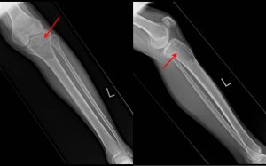
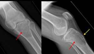
X-Ray findings:
- There is an expansile lytic lesion at proximal tibia (red arrows)
- The lesion shows well-defined margin with narrow zone of transition
- Multiple septa noted within the lesion
- Thin sclerotic border is also seen
- A cortical break is seen at anterior tibia (yellow arrow)
- No soft tissue mass. No periosteal reaction.
Biopsy done shows:
- Specimen labelled as blood consist of multiple pieces of brownish tissue measuring about 80 mm.
- Features are compatible with osteoclastic giant cells rich lesion, differentials include aneurysmal bone cyst and giant cell tumour
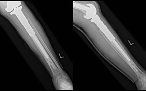
Progress of patient:
- Wide local excision with megaprosthesis fixation of proximal left tibia done
HPE findings:
- Specimen labelled as left proximal tibia consists of proximal tibia, part of fibula and surrounding soft tissue and part of overlying skin.
- Macroscopy: Cut sections of the tibia show a well-defined tumour, multiloculated, and composed of blood-filled, cystic spaces separated by tan-white, gritty septa measuring 75x80x55 mm. The fibula, surrounding soft tissue and skin are unremarkable.
- Microscopy: sections from the tumour show a well circumscribed and contains blood-filled cystic spaces separated by fibrous septa. The fibrous septa are composed of a moderately dense, cellular proliferation of bland fibroblasts with scattered multinucleated, osteoclast-type giant cells and reactive woven bone rimmed by osteoblasts. In areas, hemorrhage and hemosiderin laden macrophages are seen. The fibula, adjacent tissue and skin are unremarkable. Negative for malignancy.
- Interpretation: Aneurysmal bone cyst
Diagnosis: Aneursymal bone cyst
Discussion:
- Aneurysmal bone cysts are a rare skeletal tumours that most commonly occurs in first two decades of lifer
- They usually occur about the knee region but can occur in any axial or appendicular skeleton
- It is typically eccentrically located in the metaphysis of long bones adjacent to unfused growth plate
- Plain radiograph demonstrate a sharply demarcated expansile ostelytic lesions with thin sclerotic margins.
- CT or MRI may demonstrate fluid-fluid levels.
- Signal intensity are variables on MRI, presumably due to variable blood ages
- Bone scan shows a ‘doughnut sign’- increased uptake peripherally with a photopenic center
Recent Comments