
Case contribution: Dr Radhiana Hassan
Clinical:
- A 60 years old lady
- Presented with left breast lump.
- Mammogram and ultrasound done show a suspicious lesion in the left breast.
- Biopsy shows invasive ductal carcinoma.
- Wide local excision with axillary LN resection done
- After that she received 6 cycles of chemotherapy followed by radiotherapy
- Mammogram and ultrasound breasts done for surveillance, about 8 months after completed radiotherapy

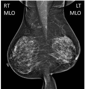
Mammogram findings:
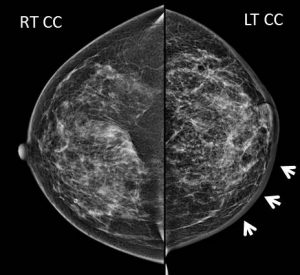
- The breasts display scattered areas of fibroglandular density (BIRADS density B).
- Left breast is smaller with associated architectural distortion in keeping with previous operation.
- Slight increased density is also observed at left breast with no dominant mass seen.
- Generalised skin thickening seen at left breast with its maximum diameter measuring about 7 mm.
- No suspicious cluster of microcalcification.
- Shotty right axillary nodes with preserved fatty hilum are observed.
Ultrasound findings:
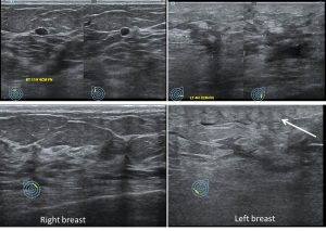
- The right breast demonstrates normal fibroglandular echogenicity and echotexture.
- A small well-defined hypoechoic lesion with posterior enhancement is seen at the right 11H measuring about 0.4 cm x 0.6 cm x 0.4 cm (AP x W x CC).
- The left breast appears oedematous particularly at the lower outer quadrant with loss of differentiation between the subcutaneous fat and fibroglandular tissues (white arrow).
- An irregular hypoechoic lesion with mild posterior shadowing is identified at left 5H, measuring about 0.4 cm x 0.5 cm x 0.5 cm (AP x W x CC). No internal vascularity is noted. This could represent scar tissue.
- No significant left axillary lymphadenopathy is seen.
Progress of patient:
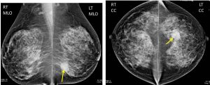
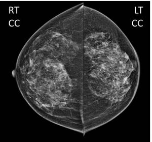
- Subsequent mammogram done after one year shows resolution of previously seen skin thickening
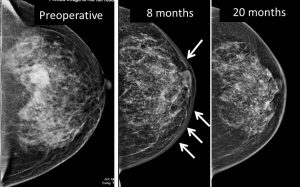
Discussion: Skin thickening post radiotherapy
- Normal skin thickness is 2 mm. Post radiation mammogram can show skin thickening with skin thickness can reach 1 cm or greater.
- Mammogram shows maximal skin thickening during the first 6 months after completion of therapy and then diminish or attain stability for many patients within 2- 3 years.
- Skin thickening after radiation is secondary to breast oedema from the damage of small vessels. Acute period of irradiation causes increased vascular permeability of breast tissue causing the breast oedema.
- It manifests as skin and trabecular thickening. It is best appreciated when compared with contra-lateral breast or with pre-treatment mammogram.
- In case of worsening skin thickening or breast oedema, differentials include lymphatic spread of cancer, obstructed venous drainage, congestive heart failure and infection. Further investigation is warranted in these cases.
Recent Comments