
Case contribution: Dr Radhiana Hassan
Clinical:
- A 57 years old male
- Presented with per rectal bleeding
- Clinical examination reveals internal haemorrhoid
- Previous history of haemorrhoidectomy
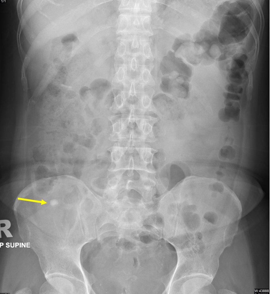
Abdominal radiograph findings:
- A rounded density seen at right iliac fossa region (yellow arrow)
- Lamilated appearance noted of this focal density.
- No dilated bowel loops.
- Normal distribution of bowel loops
- No obvious soft tissue mass lesion
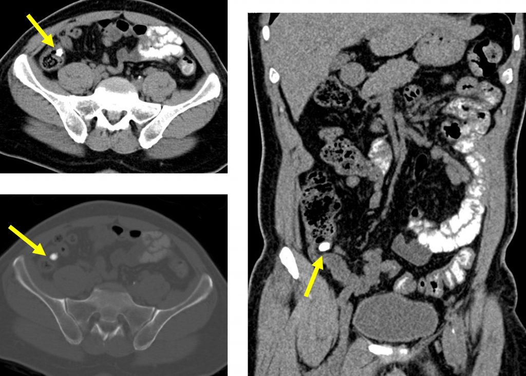
CT scan findings:
- The appendix is prominent with slightly thickened wall
- The calcification is seen within wall of proximal appendix
- No abnormal mass lesion at the bowel loops
Progress of patient:
- Colonoscopy performed with scope examination done until caecum
- There is bulging of appendicular lumen
- Biopsy was taken at terminal ileum; HPE report was normal
- Stapler granuloma at rectum from previous operation
- Patient was managed conservatively
Discussion:
- Appendicolith is a calcified deposit within the appendix
- Seen in overall 10% of patients
- May be an incidental finding on abdominal radiograph or CT scan
- Present in a large number of children with appendicitis
- A high attenuation stone at right iliac fossa on xray and CT scan
- Up to 25% show laminated appearance
- On ultrasound it will appear as lesion with acoustic shadow
Recent Comments