
Case contributions: Enam H. Moneeb, Dr Radhiana Hassan
(Extract from poster presentation “Identification of coeliac trunk variations using MDCT angiography”)
Introduction:
Variation in coeliac trunk is common and accurate knowledge of this variation is crucial before invasive procedures to avoid or reduced risk of vascular injuries.
Type of variations according to Uflacker’s classification:
| Type | Name of variation | Description |
| I | Classic trifurcation | The common hepatic artery, splenic artery and left gastric artery have a shared point of origin from the coeliac trunk |
| II | Hepatosplenic trunk | Common hepatic artery and splenic artery have shared trunk; the left gastric artery arise separately from aorta or SMA |
| III | Hepatogastric trunk | Common hepatic artery and left gastric artery have shared trunk; the splenic artery arises separately from aorta or SMA |
| IV | Hepato-spleno-mesenteric trunk | Common hepatic artery, splenic artery and SMA have shared trunk; the LGA arise separately from aorta |
| V | Gastrosplenic trunk | Left gastric artery and splenic artery have shared trunk; the common hepatic artery arise separately from aorta or SMA |
| VI | Coeliac-mesenteric trunk | Coeliac and SMA have a shared trunk |
| VII | Coeliac-colic trunk | The left or middle colic artery and the celiac have a shared trunk |
| VIII | Absent coeliac trunk | Common hepatic artery, splenic artery and left gastric arise directly from the aorta |
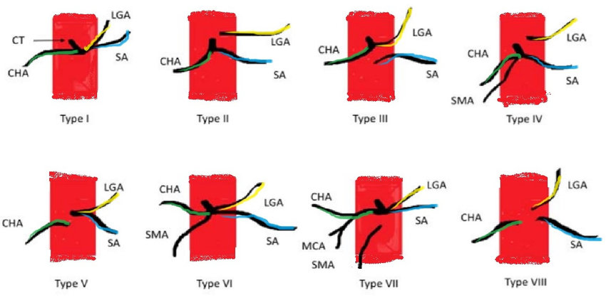
CT= coeliac trunk, CHA=common hepatic artery, SA=splenic artery, LGA=left gastric artery, SMA=superior mesenteric artery and MCA=middle colic artery
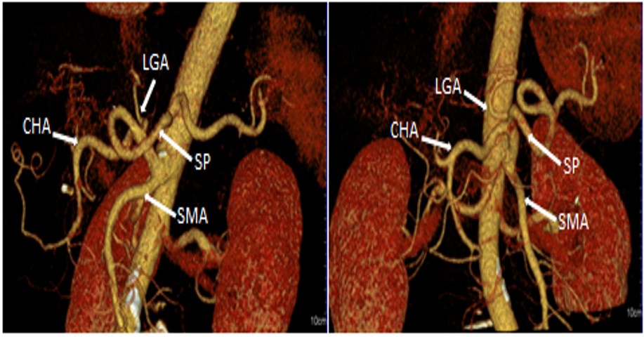
Type I: Normal trifurcation
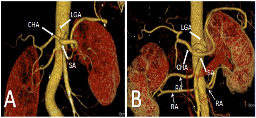
(A) Classic trifurcation: all three branches of coeliac trunk have a common origin. (B) Non-classic trifurcation where the left gastric artery may originates from any place along the coeliac trunk and in this case proximally. Notice the double renal arteries (RA).
Type II: Hepatosplenic trunk
A 3D-reconstructed image shows common hepatic artery and splenic artery as common trunk and LGA arises directly from the aorta
Type III: Hepatogastric trunk (CT image not available)
Type IV: Hepatosplenomesenteric trunk (CT image not available)
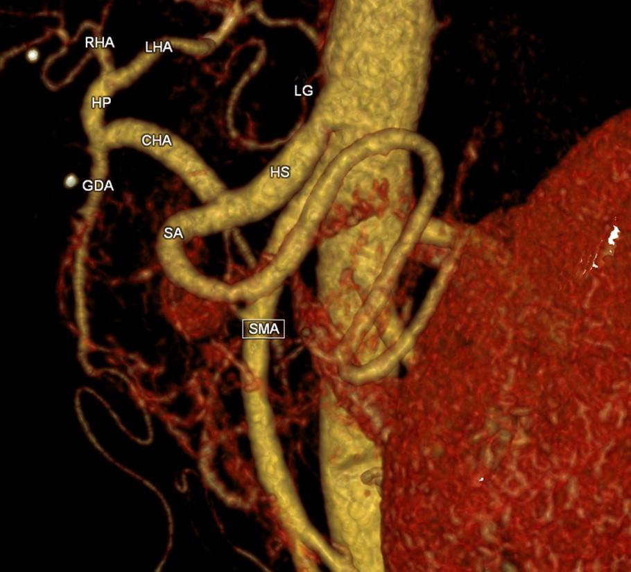
Type V: Gastrosplenic trunk
The image showed gastrosplenic trunk with left gastric artery and splenic artery as common trunk superiorly to SMA. The common hepatic artery is seen arising from SMA.
Type VI: Coeliac mesenteric trunk
The images show coeliac-mesenteric trunk where the celiac trunk and SMA have a common point of origin.
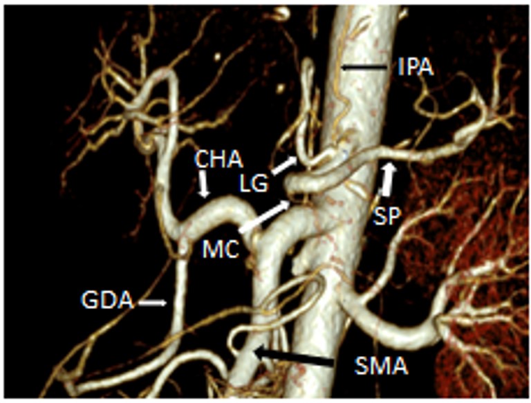
Type VII: Coeliac-colic
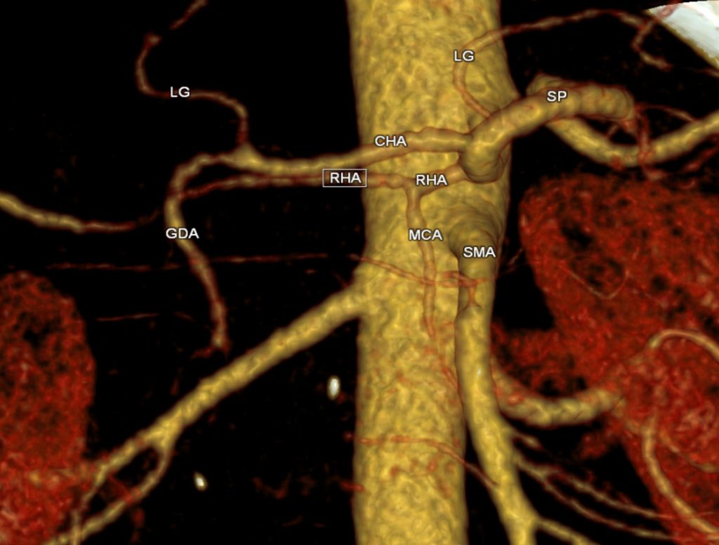
A 3D-reconstructed image shows that coeliac trunk has four branches which are common hepatic artery, left gastric artery, splenic artery and middle colic artery
Other variation: inferior phrenic arteries originates from coeliac trunk
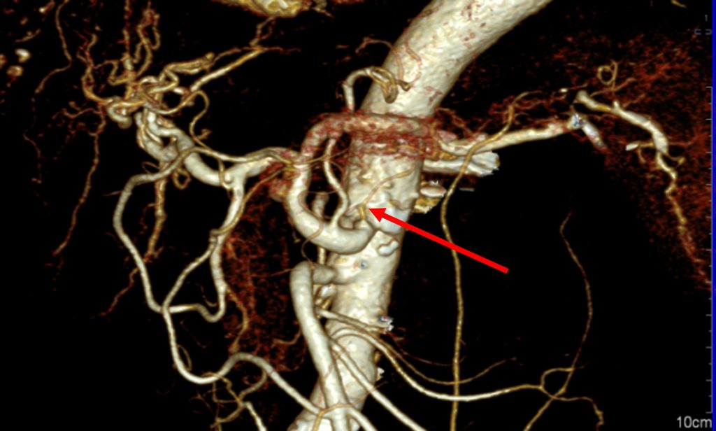
Reconstructed images of CT angiogram showing that phrenic arteries originate from a common trunk of coeliac axis. These arteries usually arise from both sides of abdominal aorta just beneath the aortic opening of diaphragm. Recent reported involvement of these arteries in the supply of hepatocellular carcinoma put more attention to these variations.
Other case: Buhler’s arc
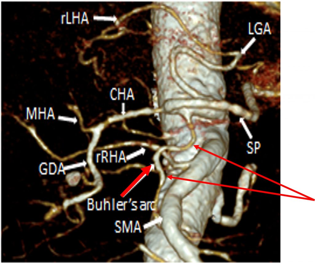
A 3D-reconstructed image shows Buhler’s arch which connects the coeliac trunk with the SMA. This has low incidence about 3% and difficult to identify.
However, this collateral vessel may open up in stenosis of SMA or coeliac trunk.
Reference:
- Uflacker, R. Atlas of vascular anatomy. An angiographic approach, 1997. Vol 81. Philadelphia. PA: Lippincott Williams
Recent Comments