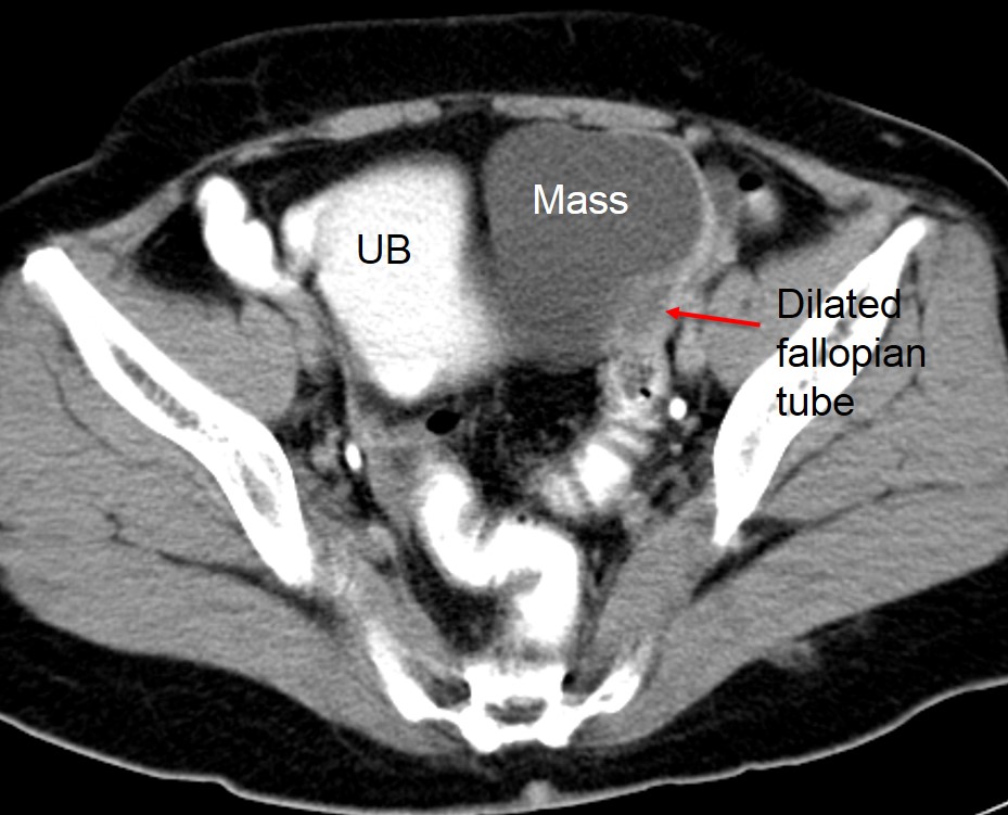
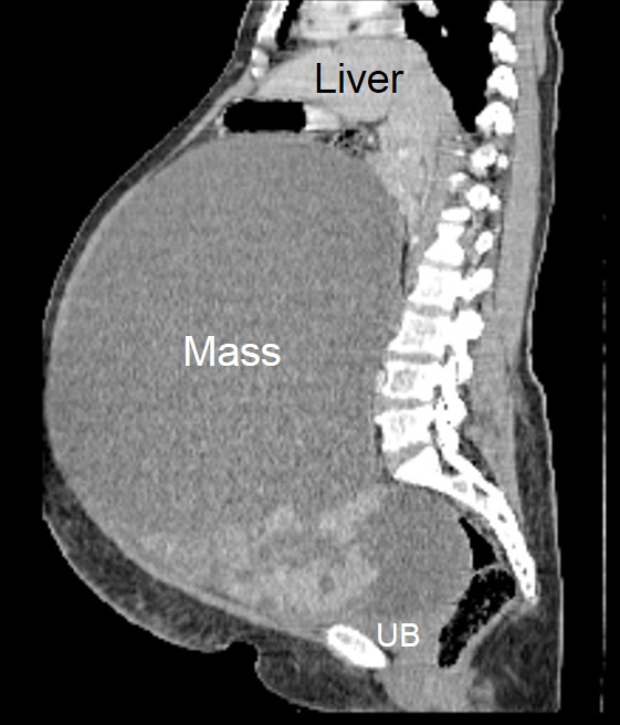
Uterine lipoleiomyoma
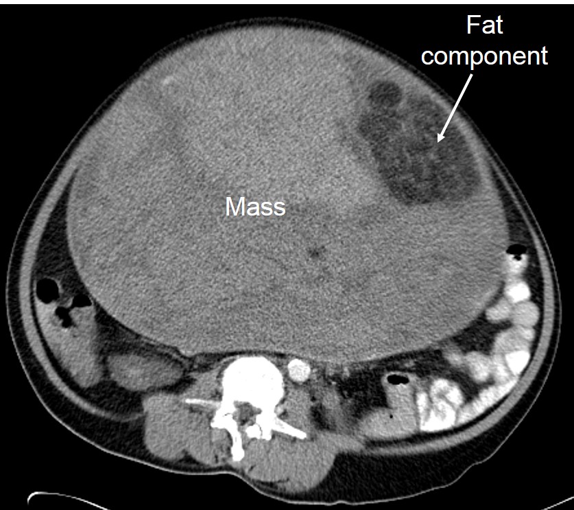
Clinical: A 53 years old lady Presented with progressive abdominal distension over few years No constitutional symptom No altered bowel habit No abnormal menses CT scan findings: A huge solid…
Twisted gangrenous ovarian mucinous cystadenoma
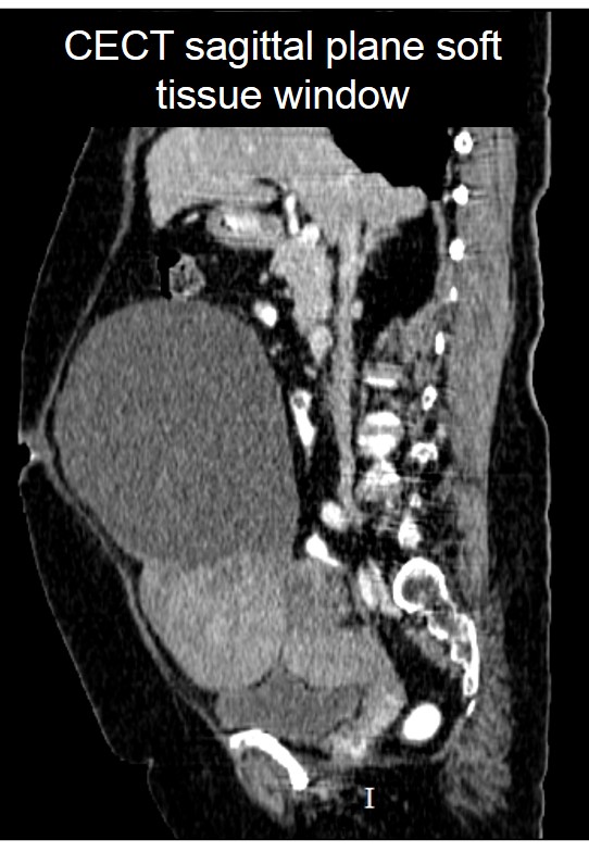
Clinical: A 61 years old lady Post menopausal Complaints of abdominal distension Gradually increase in size Presented with recent onset of abdominal discomfort No obstructive symptom CT scan findings:…
Ovarian mucinous cystadenoma 2
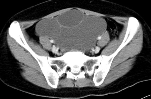
Clinical: A 24 years old lady Progressive abdominal distension No obstructive symptom CT scan findings: A cystic mass in the left pelvic region measuring 15x12x13 cm. No solid component or…
Borderline ovarian serous tumour
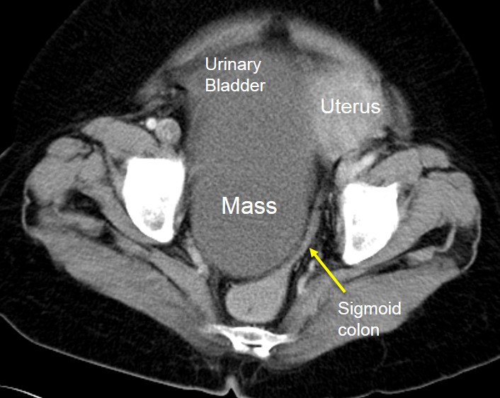
Clinical: A 60 years old lady Post menopausal Presented with chronic constipation No other obstructive symptoms CT scan findings: There is a huge cystic mass measuring 14x18x19 cm It shows…
Ovarian serous cystadenoma 2
Clinical: A 49 years old lady Incidental finding of cystic lesion in the pelvis during ultrasound done for routine medical check up. No abdominal pain. Menses are normal CT scan…
Ovarian mucinous cystadenoma of borderline malignancy
Clinical: A 52 years old lady Complaint of abdominal distension Slowly growing in size No obstructive symptoms CT scan findings: A huge abdominopelvic mass Mainly cystic in density with solid…
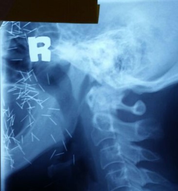
Charm needle
Clinical: A 60 years old man Presented with neck pain Radiograph of cervical spine done It showed incidental findings of charm needles at face and neck region However, clinically these…
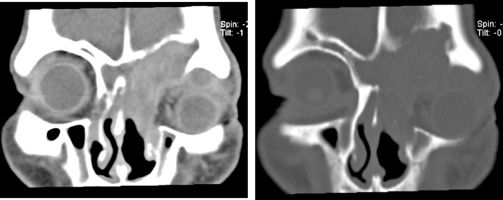
Sinonasal inverted papilloma
Clinical: A 66-year old man Underlying hypertension, otherwise no other medical problem Presented with left eye proptosis with infraorbital swelling for 10 years. Gradually increase in size Scope shows mass…
Quiz 32 Abdomen
Image: Questions: Name the examination. Name the operation that this patient has undergone. When is this examination done after the operation? Name the labelled structures. Answers: T-tube cholangiogram Cholecystectomy It…
Quiz 31 Chest
Image: Questions: Name the examination. Based on the image, which lung lobe is affected in this case? Give reasons for your answer in Question 2. How to confirm the location…
