
Clinical:
- A 24 years old lady
- No known medical illness
- Complaint of right shoulder pain for 4 years
- Intermittent pain, throbbing in nature
- Partially relieved with analgesic
- Worsened after sport activity
- No history of trauma
- No constitutional symptoms
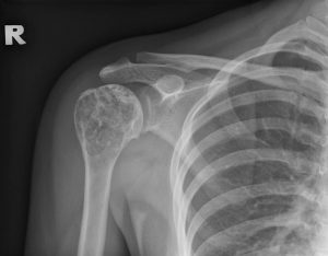
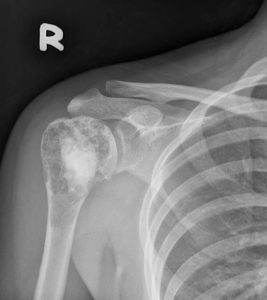
Radiographic findings:
- An expansile lytic lesion at proximal humerus
- Narrow zone of transition
- Cortical endosteal scalloping seen
- Dystrophic calcification within
- Thinning of cortex, no cortical break
- No periosteal reaction
- No surrounding soft tissue swelling
HPE findings:
- Macroscopy: specimen labeled as right proximal humerus consist of multiple strips of whitish tissue measuring 15 mm in aggregate diameter
- Microscopy: section shows strip of tumour tissues composed of multinodular to confluent architecture pattern characterized by island of hyaline cartilage encased by thin mantles of bones that are separated by marrow spaces. These islands is composed of evenly distributed and small clusters of chondrocytes. The chondrocytes are situated within sharp-edge lacunar spaces. The chondrocytes display small and round nuclei, condensed chromatin with finely granular eosinophilic and vacuolated cytoplasm. No nuclear atypia or mitosis seen. No necrosis noted.
- Interpretation: Right proximal humerus: Enchondroma
Diagnosis: Enchondroma of right proximal humerus.
Discussion:
- Enchondromas are benign cartilaginous growth in medullary cavity
- It is the most common primary benign bone tumor of hand/wrist
- It account for about 3-10% of all bone tumours and 12-24% of benign bone tumours
- Age: 10-30 years, M:F=1:1
- Usually asymptomatic painless swelling
- Usually solitary, if multiple are associated with syndromes (Ollier disease and Mafucci syndrome)
- The majority of enchondromas more frequently arise in the metaphyseal region, although they are frequently seen in the diaphysis. They only rarely are seen in the epiphysis, and a cartilaginous lesion in an epiphysis is more likely to be a chondrosarcoma.
- Enchondromas are typically located in a central or eccentric position within the medullary cavity of tubular bones:
- small tubular bones of the hands and feet; proximal phalanx most common
- large tubular bones: femur, tibia and humerus
- rare: pelvis, scapula and ribs (consider chondrosarcoma)
- Complication include pathological fracture and malignant transformation to low grade chondrosarcoma in long-bone enchondromas in 15-20% (gradual increasing pain)
- Enchondromas have a variable imaging appearance, although typically they are small lytic lesion with non-aggressive features:
- narrow zone of transition
- sharply defined margins
- +/- chondroid calcification
- expansile and may have scalloping
- should not outgrow through cortex unless with fracture
- no bone destruction, no periosteal reaction
- no soft tissue swelling
Progress of patient:
- Bone curretage and calcium sulphate insertion into humerus done.
- No complication post operatively
- Review after one year patient is well with no more pain at shoulder region
Recent Comments