
Clinical:
- A 56 years old lady
- Presented with lower abdominal fullness
- Associated with occasional vague discomfort
- Ultrasound showed a cystic pelvic mass
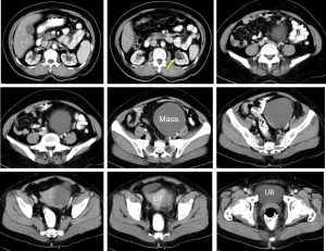
CT scan findings:
- There is a pelvic mass on the left side measuring 11x9x9 cm
- No fat component and no calcification
- Thin wall, no septae, mixed density predominantly cystic
- Normal ovaries are not identified
- No local infiltration, no ascites
- Left hydronephrosis also noted (yellow arrow)
Intra-operative findings:
- Presence of left ovarian cyst, ruptured during manipulation
- Bowel loop adhered to tumour
- Bladder high up, stuck to uterus
- Tubes bilaterally not visualised, embedded to the tumour
- No ascites, no tumour nodule on omentum
- Severe adhesion at POD
- Right ovary not visualised embedded in the mass
- Left ureter mildly dilated kinked at VUJ, right ureter normal
HPE findings:
- Macroscopy: specimen labelled as uterus, right fallopian tube, left ovarian tumour, right ovary and cervix. The left fallopian tube cannot be identified. The left fallopian tube with left ovarian tumour appear haemorrhagic with area of rupture at the posterior part of the tumour. Cut section of uterus shows normal looking uterus. Cut section of left ovarian tumour shows a multiloculated cyst with one large locule. The surface of the locule is covered by papillae which is haemorrhagic.
- Microscopy: Sections of the left ovarian tumour show it is composed of papillae and small detached cell clusters. The tumour cells have pleomorphic vesicular nuclei with prominent nucleoli and moderate amount of cytoplasm. Stromal invasion is seen. The left fallopian tube is unremarkable. The endometrium is non-secretory. Foci of endometrial glands and its stroma are seen embedded within myometrium. The cervix show nabothian cyst with no evidence of malignancy or dysplasia. No lymph nodes or any tumour deposit at omentum
- Diagnosis: Left ovarian tumour: serous adenocarcinoma with endometriosis
Diagnosis: Ovarian serous cystadenocarcinoma
Discussion:
- Ovarian serous cystadenocarcinoma is the malignant form of ovarian serous cystadenoma.
- It is the most common type of ovarian malignancy, accounts for about 25% of serous tumours.
- The incidence peaks around the 6th to 7th decades of life.
- In more than 90% of cases, there is elevated serum CA-125.
- Typical imaging features of ovarian cystadenocarcinoma include:
- cystic adnexal mass with solid component
- calcification uncommon, but can be seen about 12%
- frequently bilateral
Recent Comments