
Clinical:
- A 15 years old boy
- Involved in MVA
- On arrival in ED, GCS=15/15, BP=100/50 mmHg, PR=80 bpm
- Initial CT scan shows right renal injury
- After few days in ward patient complains of increasing pain at right lumbar region
- He also developed spiking temperature
- TWBC increased from 12.4 to 20.1
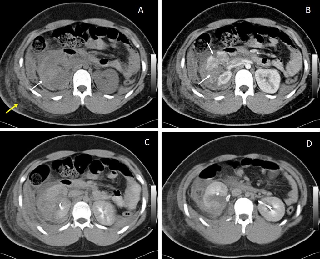
CT scan findings:
- Non contrast CT scan (A) shows hyperdense fluid at perinephric region suggestive of perinephric hematoma (white arrow). Soft tissue streakinesss and swelling also noted on the right side (yellow arrow).
- Post contrast CT scan (B) shows multiple lacerations involving the right kidney (white arrows). Some of the lacerations are deep and approaching the collection system.
- However on delayed excretory phase (C, D) there is no contrast extravasation from the collecting systems to suggest injury involving the collecting system or urinoma seen.
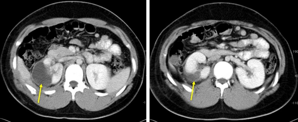
CT scan done 5 days later shows enhancing wall with streakiness of surrounding fat. Correlation with clinical information at that time, features are suggestive of infected perinephric haematoma.
Diagnosis: Infected perinephric hematoma.
Discussion:
- Secondary infection of a haematoma should be considered early if it is increasing in size or becoming more painful.
- In general, smaller abscesses are treated with IV antibiotics only and larger abscesses may require percutaneous drainage if the patient does not respond to the initial antibiotics.
- This patient was treated conservatively, responded to antibiotics and discharged well.
Recent Comments