
Case contribution: Dr Radhiana Hassan
Clinical:
- A 25 years old lady
- Presented with progressive abdominal distension
- No constitutional symptom
- No altered bowel habit
- Regular menses
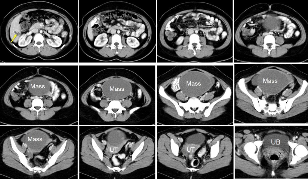
CT scan findings:
- There is well-defined predominantly cystic mass in the pelvis most likely arising from the left ovary. It measures about 13x14x10 cm.
- The mass has dense septa with irregular thickened wall at the posterior part.
- There is no calcification or fat component within it.
- The mass is compressing the uterus.
- There is a liver lesion at Segment VI with heterogenous enhancement.
- No hydronephrosis. No ascites.
HPE findings:
- Macroscopy: specimen labelled as left ovarian cyst, left fallopian tube, ovary and omentum consist of already ruptured ovarian cyst measuring 110x100x30 mm filled with jelly-like material. Cut section shows uniloculated cyst with inner surface is fully carpeted by papillary projections. The cyst wall measured 1-2 mm in thickness. No obvious solid area seen.
- Microscopy: sections from the left ovarian cyst show a tumour forming papillae. The papillae are lined by single layer to stratified ciliated epithelium. In areas, epithelial proliferation forming epithelial tufting displaying mild to moderate nuclear atypia are noted. No obvious mitosis is seen. No stromal invasion is identified. Section of the ovary shows a few cystic follicles.
- Interpretation: Left ovarian cyst: serous borderline tumour.
Diagnosis: Ovarian serous borderline tumour with liver metastasis (no biopsy of liver lesion done).
Discussion:
- Serous borderline tumour represents a group of noninvasive tumour of the ovary bridging between benign serous cystadenoma and serous carcinoma.
- They are known as a tumour of low malignant potential or as an atypical proliferative tumour.
- Serous borderline tumours account for one-fourth to one-third of the serous tumours
- They are commonly seen in younger women and usually have an excellent outcome but seldom show local recurrence
- Approximately 70% of this tumour is confined to one or both ovaries (stage I) at the time of diagnosis. The remaining tumours are found within the pelvis (stage II) or upper abdomen (stage III) and only rare cases have extended beyond the abdomen (stage IV) at the time of presentation
- They are known to spread transperitoneally. Few authors described the spread of this tumor to regional lymph nodes, supradiaphragmatic lymph nodes, and internal mammary lymph nodes.
- Rare site of metastasis include breast and brain.
Recent Comments