
Case contribution: Dr Radhiana Hassan
Clinical:
- A 47 years old man
- Presented with chronic cough
- Associated with constitutional symptoms
- Also complaint of headache
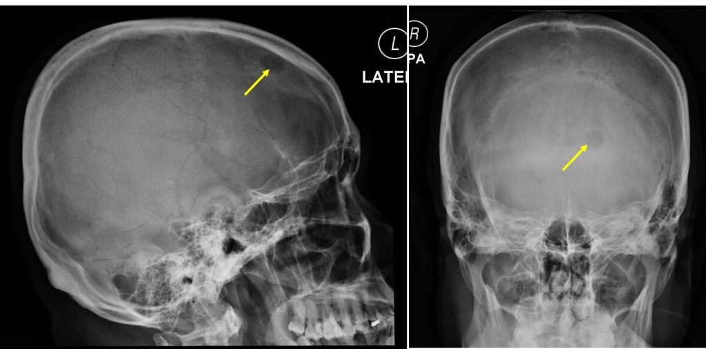
Radiographic findings:
- A rounded lytic lesion is seen at left frontal bone (arrows)
- Its outline is well delineated. No sclerotic border.
- No fluid fluid level. No internal matrix.
- No other lesion elsewhere.
- No obvious soft tissue swelling.
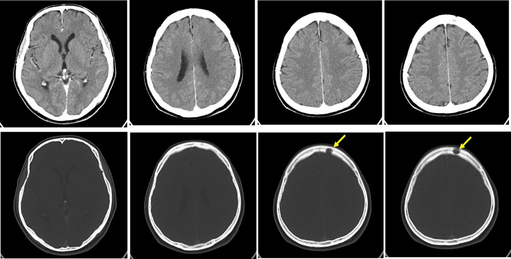
CT scan findings:
- A lytic lesion is seen at left frontal bone (arrow).
- No surrounding soft tissue mass
- No mass lesion in the brain parenchyma.
- No abnormal leptomeningeal enhancement.
- No mass effect or midline shift.
- No hydrocephalus.
Diagnosis: Lytic metastasis from lung carcinoma (HPE proven non-small cell lung ca)
Discussion:
- Bone metastasis is a common and debilitating consequence of lung cancer.
- About 30%-40% of patients with non-small cell lung cancer develop bone metastases during the course of their disease.
- The prognosis of lung cancer patients with bone metastases is poor, with a median survival time from detection of lesions measured in months.
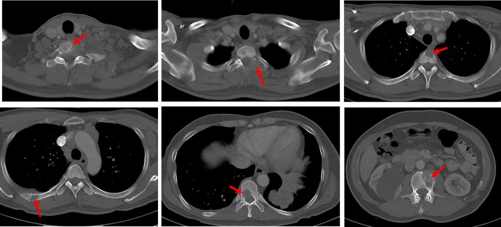
- Lung cancer bone metastases normally appear as areas of radiolucency (lytic lesions) with poor margination, no matrix and cortical destruction.
- They commonly affect the spine, ribs, pelvis and proximal long bones.
- At an early stage, bone metastases may occur easily at an axial bone through the vertebral vein system, then at appendicular bone in more advanced stages of the diseases.
- Hematogenous skull metastases can be caused by nearly all types of tumors including breast, lung, prostate, thyroid carcinoma and malignant melanoma.
- However, the most common sites for metastases from lung cancer are the upper abdomen followed by lung and pleura and lastly bone. Metastases from lung cancer involve spine and hip but skull involvement is uncommon.
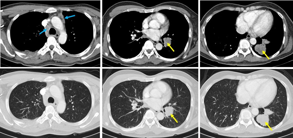
Progress of patient:
- CT scan thorax, abdomen and pelvis done shows Stage IV lung carcinoma
- Biopsy proven lung CA.
- Patient started on chemotherapy
Recent Comments