
Case contribution: Dr Radhiana Hassan
Clinical:
- A 70 years old man
- Underlying HPT, bronchial asthma, gouty arthritis and dyslipidaemia
- Presented with lower limb weakness for 5 months
- Associated with back pain and constitutional symptoms
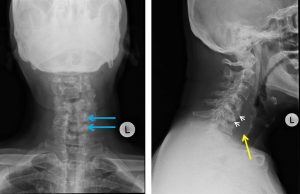
Radiographic findings:
- Loss of normal cervical lordosis. However the alignment of the vertebrae is maintained.
- Reduction and destruction of the C5 and C6 vertebral bodies (white arrows)
- Associated sclerotic change of the remnant of visualized C5 and C6 vertebrae
- The C4/C5 and C5/C6 intervertebral spaces are not seen.
- On AP view, the pedicles of the involved C4, C5, and C6 vertebra are also not visualized (blue arrows).
- The C4 vertebra body has irregular margin and appears partly sclerosed. However its height is still preserved.
- There is prevertebral soft tissue swelling from C3/C4 intervertebral disc level until the T1 vertebral body level (yellow arrow). This swelling is centered predominantly at the level of the destroyed cervical vertebral bodies. The thickest area measures about 2.7 cm.
- The rest of the visualised bones are normal.
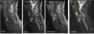
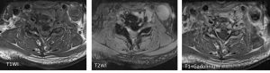
MRI findings:
- Collapse of C5 and C6 vertebral bodies (arrows). These vertebral bodies have lost their normal marrow signal and appear hypointense in T1W and T2W.
- The fragments of the collapsed C5 and C6 vertebral bodies are displaced posteriorly, indenting the anterior thecal sac and causing the narrowing of the spinal canal at their corresponding levels.
- There is no cord oedema or syrinx is observed at this level.
- The exit foramina and the exiting nerve roots are also spared.
- Irregular enhancing collection is seen in the C4/C5 intervertebral space measuring about 1.7 x 1.8 x 1.1cm (AP x W x CC). This collection is seen tracking anteriorly into the subligamentous region and extending inferiorly until T1 level (yellow arrow).
- A small suspicious looking tract is seen connecting this collection and the C6/C7 intervertebral space. The subligamentous collection measures about 0.6cm in maximal thickness.
Diagnosis: Tuberculous spondylitis (empirically treated)
Discussion:
- Tuberculous spondylitis, refers to vertebral body osteomyelitis and intervertebral diskitis due to tuberculous infection.
- It is also known as Pott disease.
- Tuberculous spondylitis can be difficult to detect in early stages because of relative preservation of the disc space. A reduction in vertebral height is often seen with the irregularity of the anterosuperior end plate being relatively early and subtle sign. Due to the subligamentous extension, there may be some irregularity of the anterior vertebral margin. This is a classical appearance with TB spondylitis.
- Empiric tuberculosis treatment is defined as the administration of TB treatment to persons being evaluated for TB who do not have laboratory evidence of TB based on high clinical suspicion.
- Empiric TB treatment strategies aim to reduce early mortality by initiating early treatment in high risk patients.
- Reassessment may rely on imaging for response
Progress of patient:
- Currently patient on maintenance phase of the therapy
- Clinically shows good response.
- Repeat MRI has not yet performed.
Recent Comments