
Clinical:
- A 55 years old man
- Underlying hypertension and DM
- Complains of left flank pain for few months
- Associated with loss of appetite and loss of weight
- No fever, no hematuria and no bowel related symptoms
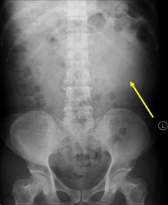
Radiographic findings:
- Soft tissue density seen at left lumbar region (yellow arrow)
- No obvious calcification is seen within the lesion
- There is peripheral displacement of bowel loops.
- Otherwise no dilatation of the bowel is seen
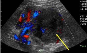
Ultrasound findings:
- A huge mass is seen in the inferior pole of left kidney
- It showed heterogenous echogenicity
- The mass is vascular with few vessels seen within it
- No hydronephrosis
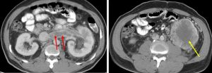
CT scan findings:
- A huge soft tissue density mass at lower pole of left kidney
- Areas of central necrosis seen (yellow arrow)
- Soft tissue density extension seen into the left renal vein (red arrows)
- No extension into the IVC
- No involvement of ipsilateral adrenal gland
Diagnosis: Renal cell carcinoma
Discussion:
- Renal cell carcinomas are the most common malignant renal tumour
- Patient usually 50-70 years of age at presentation
- Risk factors include smoking, dialysis-related cystic disease, obesity, chemotherapy agent and hypertension
- Clinical triad of macroscopic hematuria, flank pain and palpable flank mass occurs in only 10-15% of patients
- The most common sites of metastasis are lungs, bones, lymph nodes, liver, adrenals and brain
- Renal cell carcinoma has a widely varying sonographic appearance. It may appear solid or partially cystic, and may be hyper, iso, or hypoechogenic to the surrounding renal parenchyma
- On non-contrast CT the lesions are soft tissue attenuation between 20-70 HU. Larger lesions frequently have areas of necrosis.
- Approximately 30% demonstrate some calcification
- Intraluminal growth into the venous circulation, in particular, the renal vein, occurs in 4-15% of cases
Progress of patient:
- Patient was diagnosed as Stage IV disease with lung metastasis (images not shown)
- Patient refused any intervention or further treatment
- Patient died 2 months after the diagnosis
Recent Comments