
Clinical:
- A 24 years old lady
- Presented with progressive abdominal distension
- No constitutional symptoms
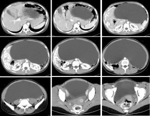
CT scan findings:
- A large cystic mass arising from pelvic region measuring 10x23x34 cm.
- No calcification, soft tissue or fat component within it
- The bowel loops are pushed peripherally. No bowel dilatation seen.
- The IVC is compressed but still patent
- Urinary bladder is also compressed by the mass.
- No hydronephrosis. No ascites
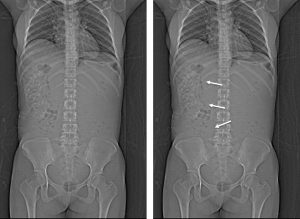
Intra-operative findings:
- Huge left ovarian cyst
- Left ovarian cytectomy done
- No spillage of cyst content intraperitoneally
- Left ovary reconstruction done
HPE findings:
- Macroscopy: specimen labelled as ovarian cyst consists of already ruptured cyst measuring 210x120x50 mm. Cut section shows a thin-walled, uniloculated cyst. No solid area seen. The cyst wall is 1-3 mm in thickness.
- Microscopy: sections show a benign, fibrocollagenous cyst wall lined by a single layer of cuboidal epithelium. There is no multilayering, atypia or mitoses seen.
- Interpretation: serous cystadenoma of left ovary
Diagnosis: Ovarian serous cystadenoma
Recent Comments