
Case contribution: Dr Radhiana Hassan
Clinical:
- A 51 years old lady
- Underlying hypertension and chronic rheumatic heart disease
- Presented with abdominal pain for one month
- Associated with post prandial vomiting
- No fever. No jaundice.
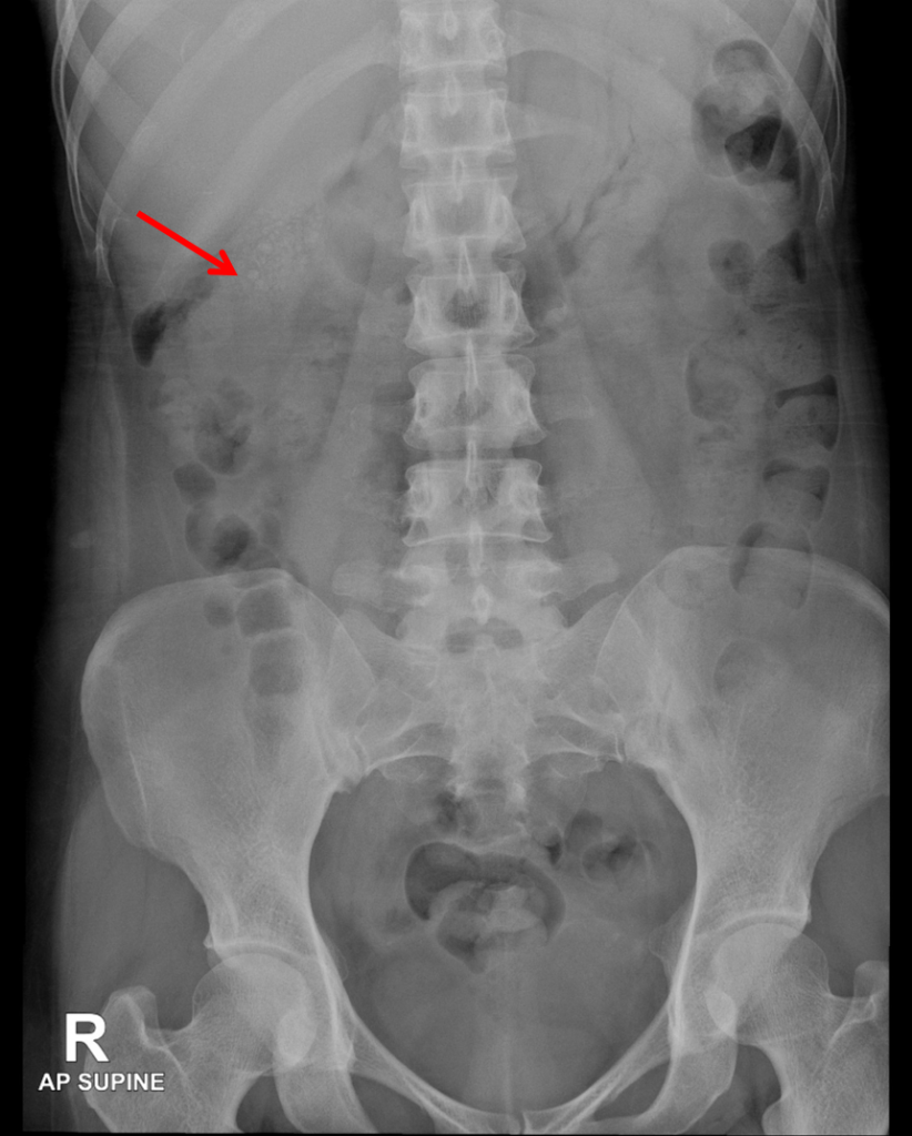
Radiographic findings:
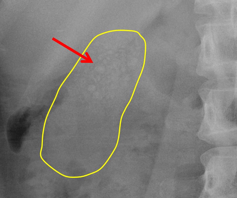
- Small stippled faint calcifications (red arrows) are observed at the lower edge of the liver, likely representing multiple gall stones.
- The largest of these stones is measured at 0.5 cm.
- Otherwise no other pathological calcification is detected.
- The bowel gas pattern is non-specific and unremarkable.
- There is no evidence of free intraperitoneal air or soft tissue mass.
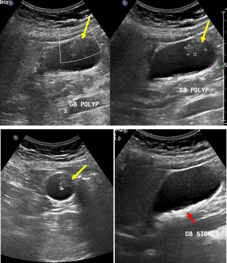
Ultrasound findings:
- Gallbladder is well distended. There is an irregular hypoechoic lesion seen arising from the anterior wall measuring about 1.3 cm x 1.8 cm. No intra-lesion vascularity detected on colour Doppler examination. This lesion does not move on patient manoeuvre.
- There are also multiple tiny calculi within the gallbladder.
- Gallbladder wall is regular and not thickened.
- No pericholecystic fluid observed. No ascites.
- No focal liver lesion is seen.
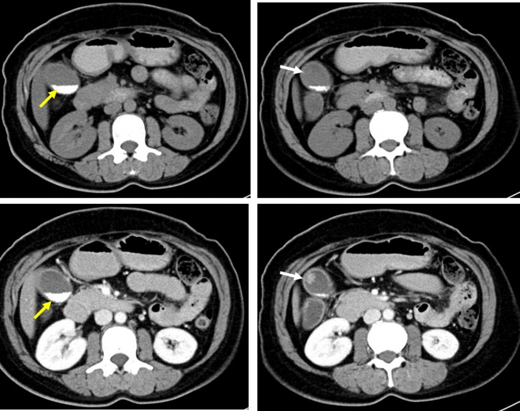
CT scan findings:
- There is a solitary polypoidal lesion seen arising from the wall of gall bladder, measuring 1.3 x 0.8 x 1.0cm (AP x W x CC). The polyp demonstrates about similar degree of enhancement with the gall bladder wall (white arrows).
- The gall bladder wall demonstrates smooth morphology, with no other polypoid lesion seen. No focal or diffuse thickening of the gall bladder wall seen.
- Numerous tiny calculi can be seen layering within the dependent aspect of the gall bladder (yellow arrows).
- No peri-cholecystic fluid detected.
HPE findings:
- Macroscopy: specimen labelled as gallbladder consists of a surgically cut opened gallbladder measuring about 78x35x15 mm. Cut section shows multiple small blackish stone within the lumen measuring 1-2 mm in widest dimension. The gall bladder wall measures 2-7 mm in thickness. No malignancy or mass seen. Representative sections are submitted: neck, body, fundus and thickened gall bladder wall.
- Microscopy: sections of gallbladder show mainly of devoid epithelial lining and replaced by granulation tissue formation with mild lymphoplasmacytic cells infiltrate. The muscular layer are hypertrophic with present of Rokitansky-Aschoff sinuses. The serosa layer is fibrotic. Negative for malignancy.
- Interpretation: chronic cholecystitis.
Diagnosis: Chronic cholecystitis.
Discussion:
- Chronic cholecystitis is prolong inflammatory condition that affects the gallbladder.
- It is almost always seen in the setting of cholelithiasis (95%) caused by intermittent obstruction of the cystic duct or infundibulum or dysmotility.
- The most commonly observed cross-sectional imaging finding are cholelithiasis and gall bladder wall thickening. The gall bladder may appear contracted or distended and pericholecystic inflammation is usually absent.
- Polyoidal appearance is not a common feature of chronic cholecystitis.
Recent Comments