
Case contribution: Dr Radhiana Hassan
Clinical:
- A 77 year old lady, underlying hypertension and neurofibromatosis 1
- Currently admitted for hypercalcaemia and investigated for colorectal cancer.
- In hospital, she had one episode of syncopal attack about one minute and spontaneously regain consciousness.
- Clinical examination shows: GCS full, power generally 5/5, sensation and reflex are normal.
- CT scan to look for ICB.
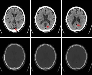
CT scan findings:
- There is no hyperdense area to suggest acute intracranial hemorrhage.
- No hypodense area to suggest cerebral infarctions.
- No midline shift or mass effect.
- Tubular hypodense lesion with fat density (mean HU -52) is noted at torcular herophili extending to the straight sinus (red arrows).
- A few similar and smaller lesions are also noted at the inferior sagittal sinus.
- Multiple nodular skin lesions in keeping with neurofibromatosis nodules.
Diagnosis: Incidental finding of fat in dural sinuses
Discussion:
- Small amounts of fat are a common incidental findings in dural sinuses
- Mostly seen in cavernous sinus, torcular herophili, the straight and transverse sinus (in decreasing order of frequency)
- These deposits represent normal adipose tissue in the dural sinuses and not lipomas
- Intracranial lipomas are seen at pericallosal, quadrigeminal cistern, suprasellar, cerebellopontine angle, sylvian fissure and choroid plexus
Recent Comments