
Case contribution: Dr Radhiana Hassan
Clinical:
- A 48 year old lady with no known medical illness
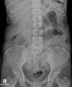
- Presented with right flank to groin pain for 1 week and dysuria
- Clinical examination shows abdomen soft and not tender
- Blood investigation: Urea 2.3, creatinine 55. Uric acid 282.
Ultrasound findings:
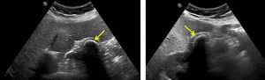
- Kidneys, ureter and urinary bladder are normal.
- No calculus in the urinary tract
- Gallbladder is partially distended. Hyperechoic lesion with posterior shadowing seen within the gallbladder and reported as big calculus.
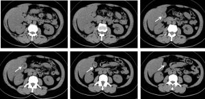
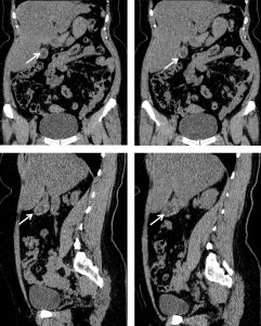
CT scan findings:
- Reverse liver (mean HU of 31) spleen (mean HU of 41) attenuation is observed in keeping with fatty liver changes.
- No focal liver lesion is detected in this plain study.
- The gallbladder is partially distended. The wall is slightly thickened. A rim calcification is seen outlining the inner wall of the gallbladder.
- No pericholecystic streakiness or collection seen.
- No urolithiasis.
Diagnosis: Incidental finding of porcelain gallbladder
Discussion:
- Porcelain gallbladder is characterized by calcification of the gallbladder wall
- It can be selective segmental mucosal calcification or complete intramural calcification with continuous band of calcium infiltrates.
- It is rare, detected in 0.06 to 0.08 percent of cholecystectomy specimens.
- There is female preponderance.
- It is associated with chronic gallbladder inflammation.
- About 95% of patients have associated gallstones.
- Patients are usually asymptomatic and diagnosis is made incidentally on imaging.
- There is associated increased in gallbladder cancer (adenocarcinoma-22%).
- Cholecystectomy has been routinely performed when porcelain gallbladder is identified.
- There is no accepted follow up interval but the annual incidence of developing gallbladder cancer is likely to be <1% per year and CT follow up is likely to be not helpful.
Recent Comments