
Case contribution: Dr Radhiana Hassan
Clinical:
- A 6 years old boy
- Presented with left eye discomfort for one month
- Parent notice abnormal growth over inner part of left eye
- Younger sibling had retinoblastoma
- Clinical examination shows ‘salmon-pink’ patch at temporal region of left eye
- Funduscopy: normal
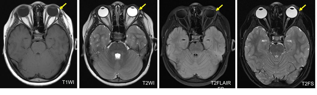
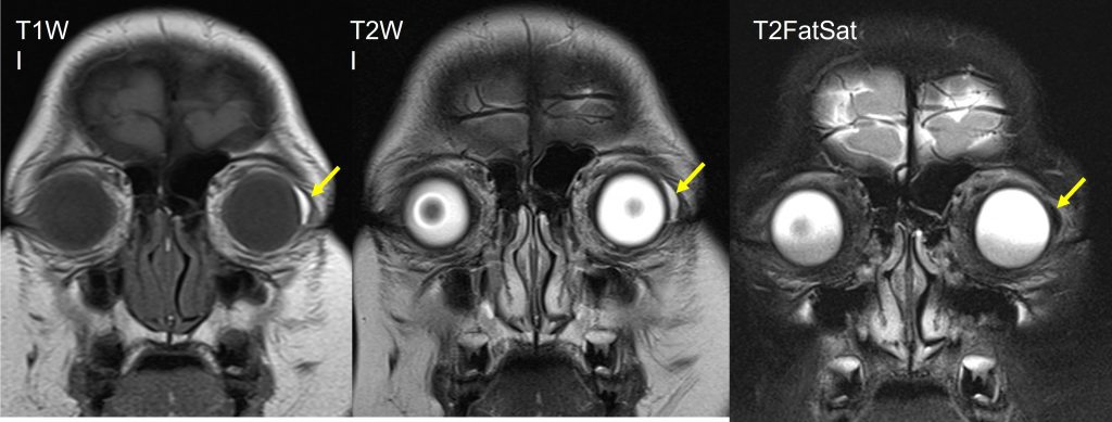
MRI findings:
- A sliver of well-defined crescent-shaped lesion abutting close at the lateral aspect of the left globe
- It is separated from the intraconal fat.
- This lesion measures about 1.0 x 0.2 x 1.0 cm (AP x W x CC).
- This lesion demonstrates signal consistent with fat i.e., hyperintense on T1 and T2 weighted images and is suppressed on fat suppression sequence.
- No blooming artefact is present to suggest hemosiderin deposit or calcification.
- Both globes and lens are normal. No proptosis.
- The extra-ocular muscles are symmetrical and normal.
- The retrobulbar fat is homogenous. No intra- or extra-conal lesion is seen.
- Both optic nerves and the optic chiasm are normal.
- The cavernous sinuses are normal.
- The superior ophthalmic veins are not dilated.
- The lacrimal glands are normal.
Diagnosis: Orbital dermolipoma (HPE proven)
Discussion:
- Orbital dermolipoma is a fat-containing epibulbar mass lesion at lateral canthal area below the superotemporal bulbar conjunvtivae.
- It is also known as epibulbar choriostoma
- It is a different entity from subconjunctival fat prolapsed
- It is congenital lesion and more commonly seen in young patients
- There is no gender predilection
- It usually present as a unilateral non-mobile firm pinkish-white or yellow mass with a straight or concave anterior margin. It not compressible and the lesion is not affected by pressure upon globe. Occasionally it may extend into the orbital fat, bulbar conjunctiva or the limbus.
- The most common differential diagnosis is subconjunctival fat prolapsed. Both lesions presented with epibulbar fatty mass in the lateral canthal area. These 2 entities are treated differently thus the importance to differentiate between the two.
- Subconjunctival fat prolapsed is an acquired lesion characterized by herniation of intraconal fat due to weakness of Tenon capsule by aging process, trauma or surgery.
- On imaging subconjunctival fat prolapsed show continuity with intraconal fat extending forward between the lateral wall of the globe medially and the lateral rectus muscle and lacrimal gland laterally.
- No enhancement post contrast. No intralesional calcification of both lesions.
- Other radiological differential diagnosis of fat-containing tumour include limbal dermoid and lipoma.
- Limbal dermoid usually seen as well-circumscribed firm solitary mass mostly located at inferotemporal or in the nasal or superior portions. Except for site it is difficult to differentiate between limbal dermoid and dermolipoma radiologically.
- Conjunctival lipoma often occurs bilaterally (50%) and mainly located in the upper fornix of the eye.
Recent Comments